Showing posts with label brain aging. Show all posts

Tuesday, September 19, 2023


A framework of biomarkers for brain aging
A framework of biomarkers for brain aging: a consensus statement by the Aging Biomarker Consortium
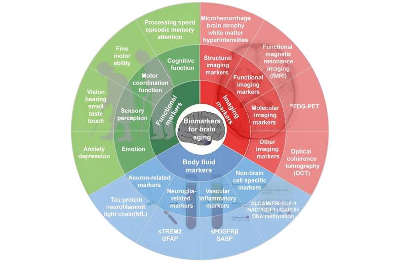
Framework of biomarkers for brain aging. Credit: Aging Biomarker Consortium

China and the world are facing severe population aging and an increasing burden of age-related diseases. Aging of the brain causes major age-related brain diseases, such as neurodegenerative diseases and stroke. Identifying biomarkers for the effective assessment of brain aging and establishing a brain aging assessment system could facilitate the development of brain aging intervention strategies and the effective prevention and treatment of aging-related brain diseases.

19 sept 2023--Thus, experts from the Aging Biomarker Consortium (ABC) have combined the latest research results and practical experience to recommend brain aging biomarkers and form an expert consensus during the seminar held on March 12, 2023 at Tongji University, aiming to provide a basis for assessing the degree of brain aging and conducting brain-aging-related research with the ultimate goal of improving the brain health of elderly individuals in both China and the world.

Biomarkers reflecting brain aging were recommended, with the aim of addressing clinical issues such as "How biologically old is the individual now?", "How fast is the individual aging?", and "How far is the individual from age-related brain diseases?" The related consensus of brain aging has been published in Life Medicine.

Brain aging involves multi-dimensional and multi-scale changes in molecules, cells, tissues, organs, the whole body, and groups of individuals. Therefore, multi-dimensional and multi-scale information needs to be integrated to accurately evaluate the state of brain aging. Brain aging biomarkers refer to markers that can accurately reflect "real age," brain structure and brain function, which can be used to determine the degree of brain aging and evaluate the effect of anti-aging interventions.

In recent years, remarkable progress has been made in exploring the biological age of the brain. Considering the accessibility and convenience of clinical procedures, this consensus screens brain aging biomarkers from the three dimensions including brain function, imaging and body fluids for reference in clinical work and follow-up studies.

Members of the ABC first identified a list of key issues related to the biomarkers for brain aging through online collaboration based on available publications and the research of the ABC members in this paper. The identified biomarkers were further discussed at a face-to-face meeting to reach a consensus.

All recommendations were fully reviewed and discussed among the members of the ABC to allow diverse perspectives and considerations for this consensus. And this consensus follows the internationally accepted conventions for expressing the level of evidence and strength of recommendations. Further validation of these markers in different age groups is needed.

More information: Yu-Juan Jia, et al, A framework of biomarkers for brain aging: a consensus statement by the Aging Biomarker Consortium, Life Medicine (2023). DOI: 10.1093/lifemedi/lnad017

Friday, June 09, 2023


Long-term type 1 diabetes associated with accelerated brain aging

Long-term type 1 diabetes associated with accelerated brain aging
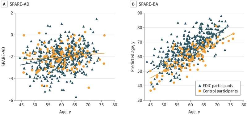
Machine Learning Indices, SPARE-AD and SPARE-BA, as a Function of Age. A, there was no significant difference observed between EDIC participants (blue) and controls without diabetes (orange). In panel B, EDIC participants showed a significant increase in predicted brain age (SPARE-BA) demonstrating more advanced brain aging patterns. SPARE-AD indicates spatial pattern for recognition-Alzheimer disease; SPARE-BA, spatial pattern for recognition-brain age. Credit: JAMA Network Open (2023). DOI: 10.1001/jamanetworkopen.2023.16182

Research led by the University of Texas Health Science Center, San Antonio, Texas, has looked into premature brain aging in individuals with type 1 diabetes.

08 Jun 2023--In the paper, "Patterns of Regional Brain Atrophy and Brain Aging in Middle- and Older-Aged Adults With Type 1 Diabetes," published in JAMA Network Open, researchers confirm a connection between type 1 diabetes and radiographic evidence accelerated brain aging.

In the study, 416 adults with type 1 diabetes who had participated in a previous observational study, Epidemiology of Diabetes Interventions and Complications (EDIC), along with 99 demographically similar adults without diabetes as a control, were assessed. The EDIC participants with type 1 diabetes had a median age of 60 years (44–74) and a median diabetes duration of 37 years (30–51).

Psychomotor and mental efficiency were evaluated using cognitive tests of verbal fluency, digit symbol substitution test, trail-making part B, and the grooved pegboard assessment. Memory scores were derived from the logical memory subtest of the Wechsler memory scale and the Wechsler digit symbol substitution test.

Greater brain age, as determined by the cognitive tests, was associated with lower psychomotor and mental efficiency among EDIC participants but was not seen among controls. The results suggest around six years of increased brain aging.

MRI scans and a machine learning program were used to calculate brain age and quantify Alzheimer's disease like atrophy. Alzheimer disease–like regional atrophy was comparable between the groups and not explicitly associated with either group. Other regions with atrophy in EDIC participants were observed, mainly in the bilateral thalamus and putamen.

The findings of this study show an increase in brain aging among individuals with type 1 diabetes without any early signs of AD-related neurodegeneration. These increases were associated with reduced cognitive performance, but according to the authors, the abnormal patterns observed in the samples were modest.

More information: Mohamad Habes et al, Patterns of Regional Brain Atrophy and Brain Aging in Middle- and Older-Aged Adults With Type 1 Diabetes, JAMA Network Open (2023). DOI: 10.1001/jamanetworkopen.2023.16182

Journal information: JAMA Network Open 

Saturday, September 03, 2022


COVID-19 infection in crucial brain regions may lead to accelerated brain aging

COVID-19 infection in crucial brain regions may lead to accelerated brain aging
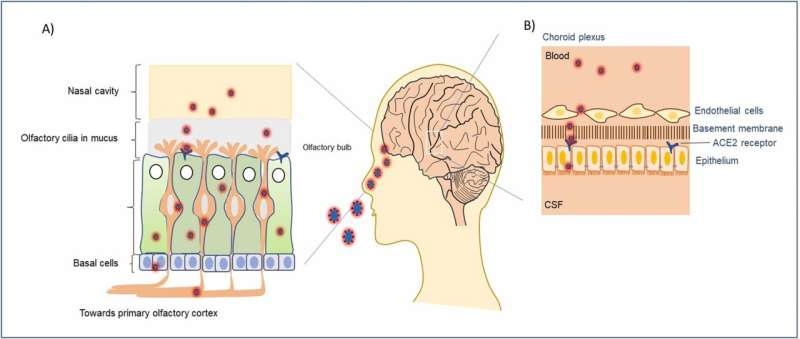
The interaction of SARS-CoV-2 with olfactory cilia for entering the central nervous system (CNS). A) Different layers of cells in the nasal cavity that contain receptors for the very first viral entry into the human host. Some of the olfactory cells express ACE2 receptors that endocytose bound viral particles, before their way to the olfactory bulb in the brain. B) Schematic of the cellular barrier between cerebrospinal fluid and circulating blood in the choroid plexus region. Both endothelial and basement epithelial cells express the ACE2 receptors facilitating the internalization of virions in the brain fluid system. Credit: Ageing Research Reviews (2022). DOI: 10.1016/j.arr.2022.101687

A new study by Houston Methodist researchers reviews the emerging insights and evidence that suggest COVID-19 infections may have both short- and long-term neurological effects. Major findings include that COVID-19 infections may predispose individuals to developing irreversible neurological conditions, may increase the likelihood of strokes and may increase the chance of developing persistent brain lesions that can lead to brain bleeding.

03 sept 2022--Led by corresponding authors Joy Mitra, Ph.D., Instructor, and Muralidhar L. Hegde, Ph.D., Professor of Neurosurgery, with the Division of DNA Repair within the Center for Neuroregeneration at the Houston Methodist Research Institute, the research team described their findings in an article titled "SARS-CoV-2 and the Central Nervous System: Emerging Insights into Hemorrhage-Associated Neurological Consequences and Therapeutic Considerations" in the journal Ageing Research Reviews.

Still a major burden on our daily lives, a great deal of research has shown that the impacts of the disease go far beyond the actual time of infection. Since the onset of the pandemic, COVID-19 has surpassed a death toll of more than 5.49 million worldwide and more than 307 million confirmed positive cases, with the U.S. accounting for almost 90 million of those cases, according to the Our World in Data website.

COVID-19 is known to invade and infect the brain, among other major organs. While a lot of research has been done to help us understand the evolution, infection and pathology of the disease, there is still a great deal that remains unclear about the long-term effects, especially on the brain.

The coronavirus infection can cause long-term and irreversible neurodegenerative diseases, particularly in the elderly and other vulnerable populations. Several brain imaging studies on COVID-19 victims and survivors have confirmed the formation of microbleed lesions in deeper brain regions related to our cognitive and memory functions. In this review study, researchers have critically evaluated the possible chronic neuropathological outcomes in aging and comorbid populations if timely therapeutic intervention is not implemented.

Microbleeds are emerging neuropathological signatures frequently identified in people suffering from chronic stress, depressive disorders, diabetes and age-associated comorbidities. Based on their earlier findings, the investigators discuss how COVID-19-induced microhemorrhagic lesions may exacerbate DNA damage in affected brain cells, resulting in neuronal senescence and activation of cell death mechanisms, which ultimately impact brain microstructure-vasculature. These pathological phenomena resemble hallmarks of neurodegenerative conditions like Alzheimer's and Parkinson's diseases and are likely to aggravate advanced-stage dementia, as well as cognitive and motor deficits.

The effects of COVID-19 infection on various aspects of the central nervous system are currently being studied. For instance, 20-30% of COVID-19 patients report a lingering psychological condition known as "brain fog" where individuals suffer from symptoms such as memory loss, difficulty in concentrating, forgetting daily activities, difficulty in selecting the right words, taking longer than usual time to complete a regular task, disoriented thought processes and emotional numbness.

More severe long-term effects analyzed in the Houston Methodist review article include predispositions for Alzheimer's, Parkinson's and related neurodegenerative diseases, as well as cardiovascular disorders due to internal bleeding and blood clotting-induced lesions in the part of the brain that regulates our respiratory system, following the COVID-19 symptoms. Additionally, cellular aging is thought to be accelerated in COVID-19 patients. A plethora of cellular stresses inhibit the virus-infected cells from undergoing their normal biological functions and let them enter into "hibernation mode" or even die completely.

The study also suggests various strategies to improve some of these long-term neuropsychiatric and neurodegenerative outcomes, as well as outlines the importance of the therapeutic regimen of the "nanozyme" in combination with various FDA-approved drugs that may prove successful to fight against this catastrophic disease.

However, given the ever-evolving nature of this field, associations like the ones described in this review show the fight against COVID-19 is far from over, say the investigators, and reinforce the message that getting vaccinated and maintaining proper hygiene are key in trying to prevent such long-term and detrimental consequences.


More information: Joy Mitra et al, SARS-CoV-2 and the central nervous system: Emerging insights into hemorrhage-associated neurological consequences and therapeutic considerations, Ageing Research Reviews (2022). DOI: 10.1016/j.arr.2022.101687
Provided by Houston Methodist 

Saturday, October 27, 2018

Landmark study sheds light on how our brains age

Landmark study sheds light on how our brains age
Credit: University of Melbourne
Two studies from a landmark 20-year Melbourne research project have shed more light on how the brain ages and what can affect the process. The results have led experts to encourage women to watch their cholesterol and blood pressure.

27 oct 2018--Two studies from a landmark 20-year Melbourne research project have shed more light on how the brain ages and what can affect the process. The results have led experts to encourage women to watch their cholesterol and blood pressure.
Both studies were published in the journal Brain Imaging and Behaviour. In the first, a person's brain volume (size) at the age of 60 predicted their memory at 70.
In this study, an MRI scan at 60 could identify those at risk of memory decline at 70, and this is supported by other international research that has identified a link between brain shrinkage and cognitive decline.
The study involved the University of Melbourne's radiology and medicine departments, Australian Catholic University's Institute for Health and Ageing and Austin Health's Aged Care Services Department.
Researchers used subjects from the population-based Women's Healthy Ageing Project, which has run at the University of Melbourne since the early 1990s. It is the first time Australian women's brain pathology has been measured alongside cognition over decades.
Sixty women had their first 3T MRI scan around the age of 59, of which 40 completed follow-up cognitive assessments over a decade. Of the 40, 23 had follow-up MRI scans.
The follow-up scans indicated that in addition to the expected age-related atrophy rate (breakdown of brain tissue), the study participants who had smaller regions of grey matter 10 years earlier were more likely to have increased rates of cognitive decline.
"This study suggests useful neuroimaging biomarkers for the prediction of cognitive decline in healthy older women," the researchers found.
The second study examined 135 Women's Healthy Ageing Project participants and found high cardiovascular risk in midlife to late life meant a higher likelihood of vascular brain damage aged over 60.
High vascular risk includes high cholesterol, low levels of the 'good cholesterol' HDL, high blood pressure, diabetes and smoking. These risks generally worsen with age.
Researchers looked at Cerebral White Matter Hyperintensity (WMH) lesions that have been identified as cerebrovascular (brain blood vessel disease) markers and are associated with increased cognitive impairment risk.
They investigated the relationship between midlife cardiovascular risk factors and late life WMH volumes two decades later, and their association with cognitive performance.
"These findings suggest intervention strategies that target major cardiovascular risk factors at midlife might be effective in reducing the development of WMH lesions and thus late life cognitive decline," the researchers found.
Study co-author Professor Cassandra Szoeke said this was the first time more than 20 years of information about Australian women had been used in this way. She said researchers looked inside the live brain using imaging data to see what impact different factors had over that time on actual brain pathology changes as well as brain function.
"In this study we showed that those women with WMH had worse cognition – the type that helps you plan, organise and get tasks done," Professor Szoeke said.
"To help reduce these risks, people should take care of their good cholesterol and blood pressure with healthy diets, activity and annual health checks."

More information: Rowa Aljondi et al. A decade of changes in brain volume and cognition, Brain Imaging and Behavior (2018). DOI: 10.1007/s11682-018-9887-z
Rowa Aljondi et al. The effect of midlife cardiovascular risk factors on white matter hyperintensity volume and cognition two decades later in normal ageing women, Brain Imaging and Behavior (2018). DOI: 10.1007/s11682-018-9970-5


Provided by University of Melbourne

Friday, September 01, 2017

Healthy glucose levels the key to a healthy ageing brain

Healthy glucose levels the key to a healthy ageing brain
New research has found blood glucose levels even at the normal range can have a significant impact on brain atrophy in ageing.

01 sept 2017--Dr Erin Walsh, lead author and post-doctoral research fellow at ANU, said the impacts of blood glucose on the brain is not limited to people with type 2 diabetes.
"People without diabetes can still have high enough blood glucose levels to have a negative health impact," said Dr Walsh from the Centre for Research on Ageing, Health and Wellbeing (CRAHW) at ANU.
"People with diabetes can have lower blood glucose levels than you might expect due to successful glycaemic management with medication, diet and exercise.
"The research suggests that maintaining healthy blood glucose levels can help promote healthy brain ageing. If you don't have diabetes it's not too early and if you do have diabetes it's not too late."
Dr Walsh said people should consider adopting healthy lifestyle habits, such as regular exercise and healthy diets.
"Having a healthy lifestyle contributes to good glycaemic control without needing a diabetes diagnosis to spur them into adopting these good habits," she said.
"It helps to keep unhealthy highly processed and sugary foods to a minimum. Also, regular physical activity every day can help, even if it is just a going for walk."
The research is part of the "Too sweet for our own good: An investigation of the effects of higher plasma glucose on cerebral health" project led by Associate Professor Nicolas Cherbuin, which is part of the longitudinal PATH through life study led by Professor Kaarin Anstey at ANU.
"The work would not be possible without being able to longitudinally explore blood glucose in members of the general public," said Dr Walsh.
The research has been published in the journal Diabetes and Metabolism.

More information: E.I. Walsh et al. Brain atrophy in ageing: Estimating effects of blood glucose levels vs. other type 2 diabetes effects, Diabetes & Metabolism (2017). DOI: 10.1016/j.diabet.2017.06.004


Provided by Australian National University

Tuesday, August 29, 2017

Dancing can reverse the signs of aging in the brain

brain

As we grow older we suffer a decline in mental and physical fitness, which can be made worse by conditions like Alzheimer's disease. A new study, published in the open-access journal Frontiers in Human Neuroscience, shows that older people who routinely partake in physical exercise can reverse the signs of aging in the brain, and dancing has the most profound effect.

29 aug 2017--"Exercise has the beneficial effect of slowing down or even counteracting age-related decline in mental and physical capacity," says Dr Kathrin Rehfeld, lead author of the study, based at the German center for Neurodegenerative Diseases, Magdeburg, Germany. "In this study, we show that two different types of physical exercise (dancing and endurance training) both increase the area of the brain that declines with age. In comparison, it was only dancing that lead to noticeable behavioral changes in terms of improved balance."
Elderly volunteers, with an average age of 68, were recruited to the study and assigned either an eighteen-month weekly course of learning dance routines, or endurance and flexibility training. Both groups showed an increase in the hippocampus region of the brain. This is important because this area can be prone to age-related decline and is affected by diseases like Alzheimer's. It also plays a key role in memory and learning, as well as keeping one's balance.
While previous research has shown that physical exercise can combat age-related brain decline, it is not known if one type of exercise can be better than another. To assess this, the exercise routines given to the volunteers differed. The traditional fitness training program conducted mainly repetitive exercises, such as cycling or Nordic walking, but the dance group were challenged with something new each week.
Dr Rehfeld explains, "We tried to provide our seniors in the dance group with constantly changing dance routines of different genres (Jazz, Square, Latin-American and Line Dance). Steps, arm-patterns, formations, speed and rhythms were changed every second week to keep them in a constant learning process. The most challenging aspect for them was to recall the routines under the pressure of time and without any cues from the instructor."
These extra challenges are thought to account for the noticeable difference in balance displayed by those participants in dancing group. Dr Rehfeld and her colleagues are building on this research to trial new fitness programs that have the potential of maximizing anti-aging effects on the brain.
"Right now, we are evaluating a new system called "Jymmin" (jamming and gymnastic). This is a sensor-based system which generates sounds (melodies, rhythm) based on physical activity. We know that dementia patients react strongly when listening to music. We want to combine the promising aspects of physical activity and active music making in a feasibility study with dementia patients."
Dr Rehfeld concludes with advice that could get us up out of our seats and dancing to our favorite beat.
"I believe that everybody would like to live an independent and healthy life, for as long as possible. Physical activity is one of the lifestyle factors that can contribute to this, counteracting several risk factors and slowing down age-related decline. I think dancing is a powerful tool to set new challenges for body and mind, especially in older age."
This study falls into a broader collection of research investigating the cognitive and neural effects of physical and cognitive activity across the lifespan.

More information: Kathrin Rehfeld et al, Dancing or Fitness Sport? The Effects of Two Training Programs on Hippocampal Plasticity and Balance Abilities in Healthy Seniors, Frontiers in Human Neuroscience (2017). DOI: 10.3389/fnhum.2017.00305


Provided by Frontiers

Thursday, August 04, 2016

Brains of overweight people 'ten years older' than lean counterparts at middle-age

Brains of overweight people 'ten years older' than lean counterparts at middle-age
Comparison of grey matter (brown) and white matter (yellow) in sex-matched subjects A (56 years, BMI 19.5) and B (50 years, BMI 43.4) Credit: Lisa Ronan
From middle-age, the brains of obese individuals display differences in white matter similar to those in lean individuals ten years their senior, according to new research led by the University of Cambridge. White matter is the tissue that connects areas of the brain and allows for information to be communicated between regions.

04 aug 2016--Our brains naturally shrink with age, but scientists are increasingly recognising that obesity - already linked to conditions such as diabetes, cancer and heart disease - may also affect the onset and progression of brain ageing; however, direct studies to support this link are lacking.
In a cross-sectional study - in other words, a study that looks at data from individuals at one point in time - researchers looked at the impact of obesity on brain structure across the adult lifespan to investigate whether obesity was associated with brain changes characteristic of ageing. The team studied data from 473 individuals between the ages of 20 and 87, recruited by the Cambridge Centre for Aging and Neuroscience. The results are published in the journal Neurobiology of Aging.
The researchers divided the data into two categories based on weight: lean and overweight. They found striking differences in the volume of white matter in the brains of overweight individuals compared with those of their leaner counterparts. Overweight individuals had a widespread reduction in white matter compared to lean people.
The team then calculated how white matter volume related to age across the two groups. They discovered that an overweight person at, say, 50 years old had a comparable white matter volume to a lean person aged 60 years, implying a difference in brain age of 10 years.
Strikingly, however, the researchers only observed these differences from middle-age onwards, suggesting that our brains may be particularly vulnerable during this period of ageing.
"As our brains age, they naturally shrink in size, but it isn't clear why people who are overweight have a greater reduction in the amount of white matter," says first author Dr Lisa Ronan from the Department of Psychiatry at the University of Cambridge, "We can only speculate on whether obesity might in some way cause these changes or whether obesity is a consequence of brain changes."
Senior author Professor Paul Fletcher, from the Department of Psychiatry, adds: "We're living in an ageing population, with increasing levels of obesity, so it's essential that we establish how these two factors might interact, since the consequences for health are potentially serious.
"The fact that we only saw these differences from middle-age onwards raises the possibility that we may be particularly vulnerable at this age. It will also be important to find out whether these changes could be reversible with weight loss, which may well be the case."
Despite the clear differences in the volume of white matter between lean and overweight individuals, the researchers found no connection between being overweight or obese and an individual's cognitive abilities, as measured using a standard test similar to an IQ test.

More information: Lisa Ronan et al, Obesity associated with increased brain-age from mid-life, Neurobiology of Aging (2016). DOI: 10.1016/j.neurobiolaging.2016.07.010


Provided by University of Cambridge

Tuesday, April 05, 2016

Exercise may slow brain aging by 10 years for older people

old person
Credit: Peter Griffin/public domain
Exercise in older people is associated with a slower rate of decline in thinking skills that occurs with aging. People who reported light to no exercise experienced a decline equal to 10 more years of aging as compared to people who reported moderate to intense exercise, according to a population-based observational study published in the March 23, 2016, online issue of Neurology, the medical journal of the American Academy of Neurology.

05 april 2016--"The number of people over the age of 65 in the United States is on the rise, meaning the public health burden of thinking and memory problems will likely grow," said study author Clinton B. Wright, MD, MS, of the University of Miami in Miami, Fla., and member of the American Academy of Neurology. "Our study showed that for older people, getting regular exercise may be protective, helping them keep their cognitive abilities longer."
For the study, researchers looked at data on 876 people enrolled in the Northern Manhattan Study who were asked how long and how often they exercised during the two weeks prior to that date. An average of seven years later, each person was given tests of memory and thinking skills and a brain MRI, and five years after that they took the memory and thinking tests again.
Of the group, 90 percent reported light exercise or no exercise. Light exercise could include activities such as walking and yoga. They were placed in the low activity group. The remaining 10 percent reported moderate to high intensity exercise, which could include activities such as running, aerobics, or calisthenics. They were placed in the high activity group.
When looking at people who had no signs of memory and thinking problems at the start of the study, researchers found that those reporting low activity levels showed a greater decline over five years compared to those with high activity levels on tests of how fast they could perform simple tasks and how many words they could remember from a list. The difference was equal to that of 10 years of aging. The difference also remained after researchers adjusted for other factors that could affect brain health, such as smoking, alcohol use, high blood pressure and body mass index.
"Physical activity is an attractive option to reduce the burden of cognitive impairment in public health because it is low cost and doesn't interfere with medications," said Wright. "Our results suggest that moderate to intense exercise may helpolder people delay aging of the brain, but more research from randomized clinical trials comparing exercise programs to more sedentary activity is needed to confirm these results."


Provided by American Academy of Neurology

Wednesday, May 06, 2015

Accelerated brain aging in type 1 diabetes related to cognitive complications

Accelerated brain aging in type 1 diabetes related to cognitive complications
Trevor Orchard. Credit: Tom Altany

The brains of people with type 1 diabetes show signs of accelerated aging that correlate with slower information processing, according to research led by the University of Pittsburgh Graduate School of Public Health.
06 may 2015--The findings indicate that clinicians should consider screening middle-aged patients with type 1 diabetes for cognitive difficulties. If progressive, these changes could influence their ability to manage their diabetes. The study, funded by the National Institutes of Health (NIH), is online and will be published in the May 19 issue of the journal Neurology.
"The severity of cognitive complications and cerebral small vessel disease—which can starve the brain of oxygen—is much more intense than we expected, but it can be measured in a clinical setting," said senior author Caterina Rosano, M.D., M.P.H., associate professor in Pitt Public Health's Department of Epidemiology. "Further study in younger patients is needed, but it stands to reason that early detection and intervention—such as controlling cardiometabolic factors and tighter glycemic control, which help prevent microvascular complications—also could reduce or delay these cognitive complications."
Type 1 diabetes usually is diagnosed in children and young adults and happens when the body does not produce insulin, a hormone that is needed to convert sugar into energy.
Dr. Rosano and her co-authors examined brain MRIs, cognitive assessments, physical exams and medical histories on 97 people with type 1 diabetes and 81 of their non-diabetic peers.
The people with type 1 diabetes were all participants in the Pittsburgh Epidemiology of Diabetes Complications Study, an ongoing investigation led by Pitt Public Health epidemiologist and study co-author Trevor Orchard, M.D., to document long-term complications of type 1 diabetes among patients diagnosed at Children's Hospital of Pittsburgh of UPMC between 1950 and 1980.
The MRIs showed that 33 percent of the people with type 1 diabetes had moderate to severe levels of  hyperintensities (markers of damage to the brain's white matter, present in normal aging and neurological disorders) compared with 7 percent of their non-diabetic counterparts.
On three cognitive tests that measure abilities such as information-processing speed, manual dexterity and verbal intelligence, the people with type 1 diabetes averaged lower scores than those without the condition.
Among only the participants with type 1 diabetes, those with greater volumes of white matter hyperintensities averaged lower cognitive scores than those with smaller volumes, though the difference was less pronounced.
The associations held even when the researchers adjusted for high blood pressure and glucose control, which are conditions that can worsen diabetes complications.
The study identified signs of nerve damage, such as numbness or tingling in extremities, as a risk factor for greater volumes of white matter hyperintensities.
"People with type 1 diabetes are living longer than ever before, and the incidence of type 1 diabetes is increasing annually," said lead author Karen A. Nunley, Ph.D., postdoctoral fellow in Pitt Public Health's neuroepidemiology program. "We must learn more about the impact of this disease as patients age. Long-term studies are needed to better detect potential issues and determine what interventions may reduce or prevent accelerated brain aging and cognitive decline."
Provided by University of Pittsburgh Schools of the Health Sciences

Monday, April 20, 2015

8 nutrients to protect the aging brain


20 april 2015--Brain health is the second most important component in maintaining a healthy lifestyle according to a 2014 AARP study. As people age they can experience a range of cognitive issues from decreased critical thinking to dementia and Alzheimer's disease. In the March issue of Food Technology published by the Institute of Food Technologists (IFT), contributing editor Linda Milo Ohr writes about eight nutrients that may help keep your brain in good shape.
  1. Cocoa Flavanols: Cocoa flavanols have been linked to improved circulation and heart health, and preliminary research shows a possible connection to memory improvement as well. A study showed  may improve the function of a specific part of the brain called the dentate gyrus, which is associated with age-related memory (Brickman, 2014).
  2. Omega-3 Fatty Acids: Omega-3 fatty acids have long been shown to contribute to good  are now playing a role in cognitive health as well. A study on mice found that omega-3 polyunsaturated fatty acid supplementation appeared to result in better object recognition memory, spatial and localizatory memory (memories that can be consciously recalled such as facts and knowledge), and adverse response retention (Cutuli, 2014). Foods rich in omega-3s include salmon, flaxseed oil, and chia seeds.
  3. Phosphatidylserine and Phosphatidic Acid: Two pilot studies showed that a combination of phosphatidylserine and phosphatidic acid can help benefit memory, mood, and cognitive function in the elderly (Lonza, 2014).
  4. Walnuts: A diet supplemented with walnuts may have a beneficial effect in reducing the risk, delaying the onset, or slowing the progression of Alzheimer's disease in mice (Muthaiyah, 2014).
  5. Citicoline: Citicoline is a natural substance found in the body's cells and helps in the development of brain tissue, which helps regulate memory and cognitive function, enhances communication between neurons, and protects neural structures from free radical damage. Clinical trials have shown citicoline supplements may help maintain normal cognitive function with aging and protect the brain from . (Kyowa Hakko USA).
  6. Choline: Choline, which is associated with liver health and women's health, also helps with the communication systems for cells within the brain and the rest of the body. Choline may also support the brain during aging and help prevent changes in brain chemistry that result in cognitive decline and failure. A major source of choline in the diet are eggs.
  7. Magnesium: Magnesium supplements are often recommended for those who experienced serious concussions. Magnesium-rich foods include avocado, soy beans, bananas and dark chocolate.
  8. Blueberries: Blueberries are known to have antioxidant and anti-inflammatory activity because they boast a high concentration of anthocyanins, a flavonoid that enhances the health-promoting quality of foods. Moderate blueberry consumption could offer neurocognitive benefits such as increased neural signaling in the brain centers.

Wednesday, April 15, 2015

The brain's aging and a new report urges ways to stay sharp

brain
Credit: Wikimedia Commons
15 april 2015--The brain ages just like the rest of a body, says a new report that urges seniors to take steps to keep sharp.
The prestigious Institute of Medicine examined what scientists know about "cognitive aging," changes in mental functioning as we get older.
This isn't a disease like Alzheimer's but a natural process—and it's not always bad. Wisdom can indeed increase with age, and years of experience can prove invaluable, stressed Dr. Dan Blazer, an emeritus professor of psychiatry at Duke University who chaired the IOM committee.
"The brain ages in all of us. But there's wide variability in the way the brain ages," Blazer said.
Staying cognitively sharp is one of the biggest concerns of seniors, with good reason. Tuesday's report warns that even subtle slowdowns can affect daily life, making seniors more vulnerable to financial scams, driving problems or other difficulties in a technology-driven world.
Indeed, while some people will experience little if any cognitive change, many older adults process information more slowly, and have more difficulty multitasking than when they were younger, the report found. What's called working memory—the brain's short-term storage—often declines with age but typically long-term memory remains intact even if it takes longer to recall someone's name.
That kind of change may not be obvious until, say, someone is faced with a complex financial decision or forced to make a transaction quickly and has trouble, Blazer said. Older adults are losing nearly $3 billion a year, directly and indirectly, to financial fraud, the report noted.
What's the difference between normal aging and cognitive decline?
"There's no clear line that we can draw here," Blazer cautioned.
Someone experiencing memory difficulty needs to be checked by a doctor, said IOM panelist Dr. Jason Karlawish of the University of Pennsylvania. With Alzheimer's, nerve cells in the brain die. With normal cognitive aging, neurons don't die—they just don't work as well, he explained.
The best advice for staying sharp as you get older: Be physically active. The sooner you start the better, but it's never too late, Blazer said.
The IOM also recommended:
—Control high blood pressure and diabetes, and don't smoke. Those are key risks for heart disease, and what's bad for your heart is bad for your brain.
—Some medications commonly taken by seniors—including certain anxiety or sleep drugs, antihistamines, bladder drugs and older antidepressants—can fog the brain, so ask about yours.
—Keep socially and intellectually active.
—Get enough sleep.
—Be careful of products that claim to improve cognitive functioning. There's no evidence that vitamins and dietary supplements like ginkgo biloba help, Blazer said. And the jury's still out on whether computer-based brain-training games do any good, he said.
The IOM also urged more research into normal cognitive aging, which has been left somewhat behind the study of diseases like Alzheimer's, and more education of doctors about their patients' risks. For example, hospitalized seniors are at increased risk for delirium—sudden confusion and agitation—that can cause lingering cognitive decline after they go home, but there are ways to prevent it.
The IOM also said government agencies and communities should consider cognitive aging as they set policies and programs. It cited a California law to protect older adults who are signing up for reverse mortgages, and a Michigan plan to improve older driver safety through such steps as adjusting traffic lights to counter glare.

Friday, March 06, 2015

Better midlife fitness may slow brain aging


People with poor physical fitness in their 40s may have lower brain volumes by the time they hit 60, an indicator of accelerated brain aging, according to new research presented at the American Heart Association EPI/Lifestyle 2015 meeting.
06 mar 2015--"Many people don't start worrying about their brain health until later in life, but this study provides more evidence that certain behaviors and risk factors in midlife may have consequences for brain aging later on," said Nicole L. Spartano, Ph.D., lead author and a postdoctoral fellow at the Boston University School of Medicine.
A subset of 1,271 participants from the Framingham Offspring Study participated in exercise treadmill testing in the 1970s, when their average age was 41. Starting in 1999, when their average age was 60, they underwent magnetic resonance imaging (MRI) of their brains as well as cognitive tests. The participants did not have heart disease or cognitive problems at the beginning of the study, and none were taking medication that alters heart rate.
In individuals with low fitness levels, the blood pressure and heart rate responses to low levels of exercise are often much higher than in individuals with better fitness.
"Small blood vessels in the brain are vulnerable to changes in blood pressure and can be damaged by these fluctuations," Spartano said. "Vascular damage in the brain can contribute to structural changes in the brain and cognitive losses. In our investigation we wanted to determine whether exaggerated blood pressure fluctuations during exercise were related to later structural changes in the brain."
The researchers found:
  • People who had a lower fitness level or greater increase in diastolic blood pressure (bottom number) or heart rate a few minutes into the low-intensity treadmill test (2.5 miles an hour) had smaller brain tissue volume later in life.
  • People who had a larger increase in diastolic blood pressure during low-intensity exercise also performed more poorly on a cognitive test for decision-making function later in life.
Poor physical fitness could be associated with accelerated brain aging.
"For every 3.4 units lower exercise capacity, every 7.1 mm Hg higher exercise diastolic blood pressure, and for every 8.3 beats/minute higher exercise heart rate in midlife, these effects are approximately equivalent to an additional 0.5 years ofbrain aging," Spartano said.
Apart from the exercise tests, a higher resting systolic blood pressure (top number) at age 40 was associated with a smaller frontal lobe volume and a greater volume of white matter hyperintensity (an indicator of loss of blood flow with aging) on the later brain MRIs.
Promotion of midlife physical fitness may be an important step towards ensuring healthy aging of the brain in the population, researchers said.
"It will be interesting to follow up with these participants in another 10 years to determine how many developed dementia, and if that may be related to their fitness or exercise blood pressure or exercise heart rate in midlife," Spartano said.
Provided by American Heart Association

Monday, December 01, 2014

Elderly brains learn, but maybe too much

Elderly brains learn, but maybe too much
Subjects were shown a sequence of letters and numbers with the distraction of dots moving in the background. Behind the numerals the dot motion became more coherent (as indicated by the arrows). Credit: Brown University
01 dec 2014--A new study led by Brown University reports that older learners retained the mental flexibility needed to learn a visual perception task but were not as good as younger people at filtering out irrelevant information.
The findings undermine the conventional wisdom that the brains of older people lack flexibility, or "plasticity," but highlight a different reason why learning may become more difficult as people age: They learn more than they need to. Researchers call this the "plasticity and stability dilemma." The new study suggests older people may indeed be facing it.
"Plasticity may be kept OK, in contrast with the view of many researchers on aging who have said that the degree of plasticity of older people gets lower," said Takeo Watanabe, the Fred M. Seed Professor at Brown University, corresponding author of the study in Current Biology. "However, we have found that the stability is problematic. Our learning and memory capability is limited. You don't want older, existing important information that is already stored to be replaced with trivial information."
Numerals, not dots
To conduct the study, Watanabe and his team enrolled a group of 10 people between 67 and 79 years old and another group of 10 people ages 19 to 30 for an experiment. Over a nine-day period, they trained on a simple visual exercise: Shown a quick sequence of six symbols - four letters and two numerals - volunteers were asked to report the numerals they saw. Their performance on a test at the end of training was compared to their score on a pre-test.
The volunteers were explicitly instructed to only bother with spotting the two numerals, but each symbol they saw had a background of moving dots. Unbeknownst to the subjects, those dots would move with varying degrees of cohesiveness of direction. In the pre- and post-tests the researchers also asked the volunteers to report the direction of dot movement when they saw the numerals.
The results of the testing were telling. Older people improved as much as younger people on the relevant task of identifying the two numerals.
"These results indicate that older subjects as well as younger subjects showed significant amounts of task-relevant learning," the authors wrote. "No evidence was obtained that indicates that older individuals have a problem with plasticity."
Last week, in fact, Watanabe and colleagues published a study showing that plasticity during visual learning occurs in older people as well as younger ones, but it is manifest differently in the brains of the different groups.
But in this study when it came to the irrelevant skill of discerning the prevailing direction of dot movement, older people learned that, too, even when it was at its most obvious. Younger people, meanwhile, only showed improvement on discerning movement when it was insidiously subtle. If it was clear, they recognized it and filtered it out.
The idea that the most obvious signals were the most easily filtered, suggested that the difference between older and younger learners was a matter of attention.
The researchers therefore subjected the volunteers to another test for the ability to find a relevant stimulus amid a number of distractors. Older people did notably worse than younger ones, adding evidence that the attentional systems for filtering out irrelevant stimuli were indeed weaker in older learners. Importantly, the poorer an older subject was at the ability to filter out irrelevant stimuli, the more irrelevant stimuli the subject learned.
Watanabe said the finding is not necessarily discouraging news. Perhaps filtering can be improved with some kind of training.
"The hope is that maybe what older people need to do is to learn a skill to avoid learning what is not necessary," he said.
More information: Current Biology, Chang et al.: "Age-related declines of stability in visual perceptual learning" www.cell.com/current-biology/a… 0960-9822(14)01350-5
Provided by Brown University

Monday, January 19, 2009

Canada-US scientists discover gene responsible for brain's aging

Universite de Montreal, Maisonneuve-Rosemont Hospital and Lawrence Berkeley National Laboratory publish findings in the Journal of Neuroscience

Montreal, 19 jan 2009– Will scientists one day be able to slow the aging of the brain and prevent diseases such as Alzheimer's and Parkinson's? Absolutely – once the genetic coding associated with neuronal degeneration has been unraveled.

According to a new study published in The Journal of Neuroscience, a research team from the Université de Montréal, Maisonneuve-Rosemont Hospital and Lawrence Berkeley National Laboratory has taken a giant step in this direction by identifying a gene that controls the normal and pathological aging of neurons in the central nervous system: Bmi1.

The primary risk factor for diseases such as macular degeneration, Parkinson's and Alzheimer's is age. Although many researchers have sought to better understand the genetics and pathophysiology of these diseases, few studies have focused on the basic molecular mechanisms that control neuronal aging.

Dr. Gilbert Bernier, of the Université de Montréal and Maisonneuve-Rosemont Hospital, led a team that identified a mutation in mice that dramatically accelerates the process of aging in the brain and the eye. The new study reveals that neurons in the retina and cerebral cortex require a gene called Bmi1 to prevent activation of the p53 pathway and the accumulation of free radicals.

"Overall, we have now established that the Bmi1 gene is a direct regulator of cell aging in brain and retinal neurons of mammals through its action on the defense mechanisms against free radicals," says Dr. Bernier.

###

About the study:

The article entitled "The Polycomb Group Gene Bmi1 Regulates Antioxidant Defenses in Neurons by Repressing p53 Pro-Oxidant Activity," published in The Journal of Neuroscience, is the work of Dr. Gilbert Bernier in collaboration with Wassim Chatoo, Mohammed Abdouh, Jocelyn David, Marie-Pier Champagne, José Ferreira from the Université de Montréal and Maisonneuve-Rosemont Hospital with Francis Rodier from the Lawrence Berkeley National Laboratory, San Francisco, U.S.

On the Web:

About the Université de Montréal: www.umontreal.ca.
About the Hôpital Maisonneuve-Rosemont: www.maisonneuve-rosemont.org
About the Lawrence Berkeley National Laboratory: www.lbl.gov