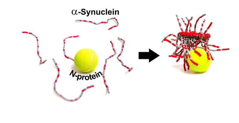
The SARS-CoV-2 N-protein can interact with α-synuclein in the test tube and help it form amyloid fibrils, a hallmark of Parkinson's disease. Credit: Adapted from ACS Chemical Neuroscience 2021, DOI: 10.1021/acschemneuro.1c00666
Case reports of relatively young COVID-19 patients who developed Parkinson's disease within weeks of contracting the virus have led scientists to wonder if there could be a link between the two conditions. Now, researchers reporting in ACS Chemical Neuroscience have shown that, at least in the test tube, the SARS-CoV-2 N-protein interacts with a neuronal protein called α-synuclein and speeds the formation of amyloid fibrils, pathological protein bundles that have been implicated in Parkinson's disease.
26 dec 2021--In addition to respiratory symptoms, SARS-CoV-2 can cause neurological problems, such as loss of smell, headaches and "brain fog." However, whether these symptoms are caused by the virus entering the brain, or whether the symptoms are instead caused by chemical signals released in the brain by the immune system in response to the virus, is still controversial. In Parkinson's disease, aproteincalled α-synuclein forms abnormal amyloid fibrils, leading to the death of dopamine-producing neurons in the brain. Interestingly, loss of smell is a common premotor symptom in Parkinson's disease. This fact, as well as case reports of Parkinson's in COVID-19 patients, made Christian Blum, Mireille Claessens and colleagues wonder whether protein components of SARS-CoV-2 could trigger the aggregation of α-synuclein into amyloid. They chose to study the two most abundant proteins of the virus: the spike (S-) protein that helps SARS-CoV-2 enter cells, and the nucleocapsid (N-) protein that encapsulates the RNA genome inside the virus.
In test tube experiments, the researchers used a fluorescent probe that binds amyloid fibrils to show that, in the absence of the coronavirus proteins, α-synuclein required more than 240 hours to aggregate into fibrils. Adding the S-protein had no effect, but the N-protein decreased the aggregation time to less than 24 hours. In other experiments, the team showed that the N- and α-synuclein proteins interact directly, in part through their opposite electrostatic charges, with at least 3–4 copies of α-synuclein bound to each N-protein. Next, the researchers injected N-protein and fluorescently labeled α-synuclein into a cell model of Parkinson's disease, using a similar concentration of N-protein as would be expected inside a SARS-CoV-2-infected cell. Compared to control cells with only α-synuclein injected, about twice as many cells died upon injection of both proteins. Also, the distribution of α-synuclein was altered in cells co-injected with both proteins, and elongated structures were observed, although the researchers could not confirm that they were amyloid. It's unknown whether these interactions also occur within neurons of the human brain, but if so, they could help explain the possible link between COVID-19 infection and Parkinson's disease, the researchers say.
More information: Slav A. Semerdzhiev et al, Interactions between SARS-CoV-2 N-Protein and α-Synuclein Accelerate Amyloid Formation, ACS Chemical Neuroscience (2021). DOI: 10.1021/acschemneuro.1c00666
Provided by American Chemical Society
Saturday, January 02, 2021
Unique susceptibility to unique Sars-CoV-2 variants and vaccines
by John Hewitt , Medical Xpress
Credit: Wikipedia
Individuals with different genetic variants in their immune system components often have very different immune responses to Sars-CoV-2. They also will have different responses to vaccines. By the same token, newly emerged variants in Sars-Cov-2 can elicit different immune responses in identical immune systems. In the larger reality we are now dealing, potential variation in all the above must be simultaneously considered.
02 Jan 2021--We recently discussed several sources of newly uncovered variation inimmune genesthat control susceptibility to Sars. For example, modern humans with throwback versions of theneanderthalgene DPP4, or the spike protein cleaving protease TMPRSS2 appear to be at high risk for severe Covid. Other variants, like a highly-expressedTMEM1Bgene common in East Asians, or in genes of the heparin sulfate synthesis pathway, help explain the disproportionate severity of COVID-19 in some populations
Two papers, one recently published in Nature Immunology, and the other in Science, now extend the emerging gene list to include variations in the structure of the antibodies that are elicited by virus or vaccine. These variations include a particular kind of posttranslational modification of the anti-RBD (receptor binding domain) of the abundant IgG1 subclass known as afucosylation. What this essentially means is that for one reason or another, the afucosylated antibody is missing the addition of a fucose sugar molecule at a key structural location.
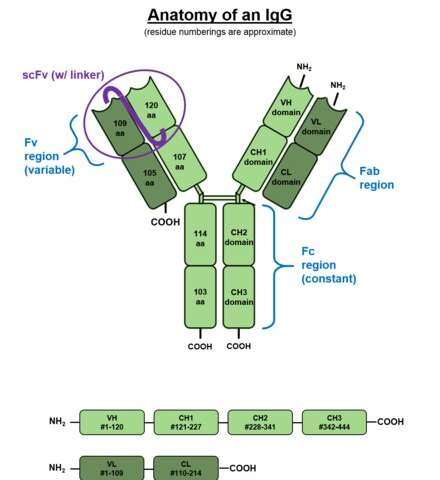
To better visualize the effect of this situation consider the above picture of the standard issue IgG antibody molecule. The variable portion, the arms of the Y shaped molecule, is known as the Fab region and it contains the epitopes that interact with the virus spike protein. The singular base region contains the constant Fc fragment, and incidentally, the glycosylated adornment hanging off of position N297 that potentially contains the fucose. In an almost fractal-like idiosyncrasy, this glycoform piece of kit also has a similar biantennary structure to the parent antibody, only on a much smaller scale, and with various sugars instead of amino acids. The fucose itself hangs off the stem portion of the aggregate glycoform, much as these glycans in turn hang on the antibody.
For anyone familiar with a related type of posttranslational modification known as ubiquitination, the N-glycan linkages form a kind of a code, much like the so-called ubiquitin code. Whereas the ubiquitin code consists of linear and branching subunits of variously phosphorylated or acetylated ubuitins, SUMOs, or NEDDs, the N-glycan code is composed of a heptasaccharide core which can be further extended with core fucose, terminal galactose (Gal), terminal sialic acid (Sia), and bisecting GlcNAc through selective enzymatic glycosylation reactions.
What the authors of these new papers found is that patients with severe forms of Sars-CoV-2 had increased likelihood of IgG1 antibodies with afucosylated Fc glycans, particularly in males. The net result of afucosylation proved to be enhanced interaction and binding with the immune activating Fcγ receptor, FcγRIIIA. This subsequently increases production of cytokines like interleukin-6 and TNF by monocytes. It should be noted that the canonical Fc receptors for human IgG include both activating (FcγRI, FcγRIIA, FcγRIIC, FcγRIIIA, and FcγRIIIB) and inhibitory (FcγRIIB) receptors. Most immune effector cells coexpress both activating and inhibiting FcγRs and therefore the net outcome of IgG interactions can often be predicted as the ratio of the binding affinities of a specific activating to inhibiting IgG receptors.
The way the authors figured this kind of stuff out was to first isolate the relevant IgG from a patient's serum using protein purification and tryptic digestion. Then nanoscale liquid chromatography coupled to tandem mass spectrometry is generally used to characterize the sites of potential glycosylation. But what the authors really wanted to get at was a more quantitative description of how much receptor binding is affected by afucosylation. To do this, various tried-and-true biophysical techniques including Surface Plasmon Resonance (SPR), Isothermal Titration Calorimetry (ITC), Microscale Thermophoresis (MST), and Biolayer Interferometry (BLI) are now available.
The authors of the Nature paper choose to use Biolayer Interferometry, which yielded graphs for binding (in nm units) as a function of time from which they could derive kinetic constants. BLI is an optical, label-free technique that analyzes the interference pattern of white light reflected from two surfaces. One is a layer of immobilized protein on the biosensor tip while the other is an internal reference. When a ligand immobilized on the biosensor tip surface binds to an analyte in solution there is a shift in the interference pattern that can be measured in real-time.
Afucosylated IgG was found to have a 20–40-fold increase in affinity to FcγRIIIa. This helps explain observed shifts in the front-line lung scavengers expressing FcγRIIIA, namely the alveolar macrophages, into an activated state of antibody-dependent cellular phagocytosis. While lowered Fc fucosylation is seen in the anti-Spike responses of the ARDS patients, other research has found that high levels of fucosylation are found in several kinds of cancer. In attempting to generate high-titer convalescent immunoglobulin treatments, it is likely important that plasma enriched in fucosylated anti-SARS-CoV-2 antibodies should be used. Although afucosylated IgG formed against viruses generally mediate stronger FcγRIIIa responses, they will often amplify cytokine storm and immune pathology.
While variants in the many genes that help build glycan chains, like for example the fucosyltransferase FUT2, might be expected to play a role in patient responses, perhaps an ever more pressing concern is differences in the antibody response to vaccines. In a brief spate of good news, the actual sequence of the new Pfizer mRNA vaccine was generously released unto the masses. The quickest to respond and attempt to decode this revelation was a guy named Bert Hubert, who quickly broke it down for us.
The main difference in the vaccine code is that uracil has been replaced by 1-methyl-3'-pseudouridylyl, which is labelled as Ψ. Although Ψ does not arouse the ire of our immune systems, it is still accepted as a normal uracil by the translation, transcription, and replication machinery of the cell. The other curious features are the substitutions of two prolines to stabilize the spike structure, and the deployment of special 5' and 3' untranslated regions before and after the main spike sequence. Bert has promised to release a second deep dive into the sequence in a few days, which is no doubt anxiously awaited by many.
More information: Saborni Chakraborty et al. Proinflammatory IgG Fc structures in patients with severe COVID-19, Nature Immunology (2020). DOI: 10.1038/s41590-020-00828-7
Mads Delbo Larsen et al. Afucosylated IgG characterizes enveloped viral responses and correlates with COVID-19 severity, Science (2020). DOI: 10.1126/science.abc8378
Monday, November 02, 2020
Coronavirus mutation may have made it more contagious: study
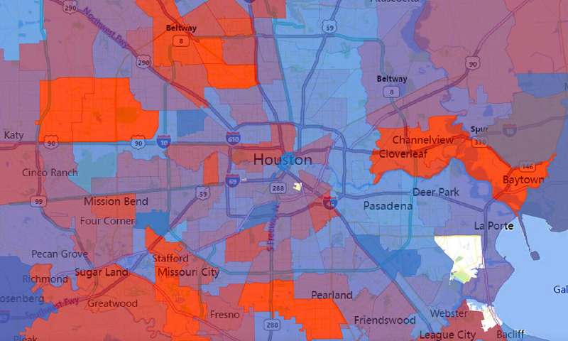
The number of virus strains present in each zip code in Houston during the second wave of COVID-19 cases in summer 2020. Number of strains is represented by a spectrum of colors from blue (0 strains) to red (50 strains). Credit: Houston Methodist/University of Texas at Austin.
A study involving more than 5,000 COVID-19 patients in Houston finds that the virus that causes the disease is accumulating genetic mutations, one of which may have made it more contagious. According to the paper published in the peer-reviewed journal mBIO, that mutation, called D614G, is located in the spike protein that pries open our cells for viral entry. It's the largest peer-reviewed study of SARS-CoV-2 genome sequences in one metropolitan region of the U.S. to date.
02 november 2020--The paper shows "the virus is mutating due to a combination of neutral drift—which just means random genetic changes that don't help or hurt the virus—and pressure from our immune systems," said Ilya Finkelstein, associate professor of molecular biosciences at The University of Texas at Austin and co-author of the study. The study was carried out by scientists at Houston Methodist Hospital, UT Austin and elsewhere.
During the initial wave of the pandemic, 71% of the novel coronaviruses identified in patients in Houston had this mutation. When the second wave of the outbreak hit Houston during the summer, this variant had leaped to 99.9% prevalence. This mirrors a trend observed around the world. A study published in July based on more than 28,000 genome sequences found that variants carrying the D614G mutation became the globally dominant form of SARS-CoV-2 in about a month. SARS-CoV-2 is the coronavirus that causes COVID-19.
So why did strains containing this mutation outcompete those that didn't have it?
Perhaps they're more contagious. A study of more than 25,000 genome sequences in the U.K. found that viruses with the mutation tended to transmit slightly faster than those without it and caused larger clusters of infections. Natural selection would favor strains of the virus that transmit more easily. But not all scientists are convinced. Some have suggested another explanation, called "founder's effects." In that scenario, the D614G mutation might have been more common in the first viruses to arrive in Europe and North America, essentially giving them a head start on other strains.
The spike protein is also continuing to accumulate additional mutations of unknown significance. The Houston Methodist-UT Austin team also showed in lab experiments that at least one such mutation allows spike to evade a neutralizing antibody that humans naturally produce to fight SARS-CoV-2 infections. This may allow that variant of the virus to more easily slip past our immune systems. Although it is not clear yet whether that translates into it also being more easily transmitted between individuals.
The good news is that this mutation is rare and does not appear to make the disease more severe for infected patients. According to Finkelstein, the group did not see viruses that have learned to evade first-generation vaccines and therapeutic antibody formulations.
"The virus continues to mutate as it rips through the world," Finkelstein said. "Real-time surveillance efforts like our study will ensure that global vaccines and therapeutics are always one step ahead."
The scientists noted a total of 285 mutations across thousands of infections, although most don't appear to have a significant effect on how severe the disease is. Ongoing studies are continuing to surveil the third wave of COVID-19 patients and to characterize how the virus is adapting to neutralizing antibodies that are produced by our immune systems. Each new infection is a roll of the dice, an additional chance to develop more dangerous mutations.
"We have given this virus a lot of chances," lead author James Musser of Houston Methodist told The Washington Post. "There is a huge population size out there right now."
Several other UT Austin authors contributed to the work: visiting scholar Jimmy Gollihar, associate professor of molecular biosciences Jason S. McLellan and graduate students Chia-Wei Chou, Kamyab Javanmardi and Hung-Che Kuo.
The UT Austin team tested different genetic variants of the virus's spike protein, the part that allows it to infect host cells, to measure the protein's stability and to see how well it binds to a receptor on host cells and to neutralizing antibodies. Earlier in the year, McLellan and his team at UT Austin, in collaboration with researchers at the National Institutes of Health, developed the first 3-D map of the coronavirus spike protein for an innovation that now factors into several leading vaccine candidates' designs.
The researchers found that SARS-CoV-2 was introduced to the Houston area many times, independently, from diverse geographic regions, with virus strains from Europe, Asia, South America and elsewhere in the United States. There was widespread community dissemination soon after COVID-19 cases were reported in Houston.
An earlier version of the paper was posted last month to the preprint server medRxiv.
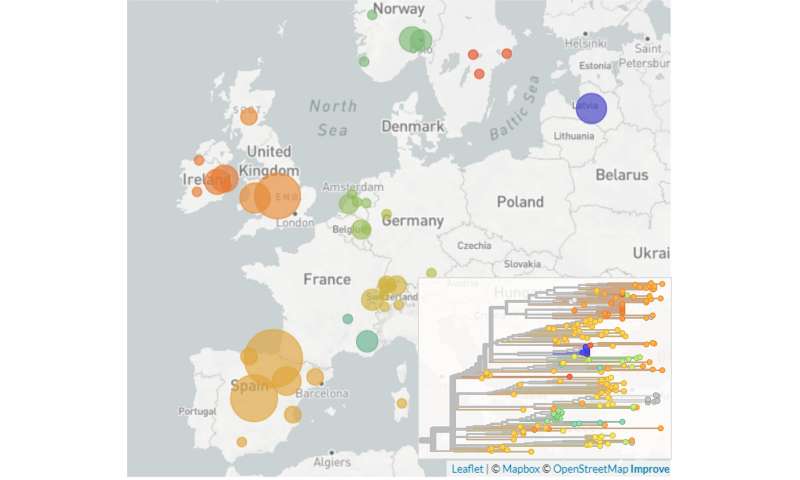
Spread of a novel SARS-CoV-2 variant across Europe in summer 2020. Credit: University of Basel
Researchers from Basel and Spain have identified a novel SARS-CoV-2 variant that has spread widely across Europe in recent months, according to an un-peer-reviewed preprint released this week. While there is no evidence of this variant being more dangerous, its spread may give insights into the efficacy of travel policies adopted by European countries during the summer.
02 november 2020--In Europe alone, hundreds of different variants of the newcoronavirusSARS-CoV-2 are currently circulating, distinguished by mutations in their genomes. However, only very few of these variants have spread as successfully and become as prevalent as the newly identified variant, named 20A.EU1.
The researchers at the University of Basel, ETH Zürich in Basel and the SeqCOVID-Spain consortium analyzed and compared virus genome sequences collected from COVID-19 patients all across Europe to trace the evolution and spread of the pathogen (see box). Their analysis suggests that the variant originated in Spain during the summer. The earliest evidence of the new variant is linked to a super-spreading event among agricultural workers in the north-east of Spain. The variant moved into the local population, expanding quickly across the country, and now accounts for almost 80% of the sequences from Spain.
"It is important to note that there is currently no evidence the new variant's spread is due to a mutation that increases transmission or impacts clinical outcome," stresses Dr. Emma Hodcroft of the University of Basel, lead author of the study. The researchers believe that the variant's expansion was facilitated by loosening travel restrictions and social distancing measures in summer.
Similar pattern as in spring in Spain
"We see a similar pattern with this variant in Spain as we did in the spring," advises Professor Iñaki Comas, co-author on the paper and head of the SeqCOVID-Spain consortium. "One variant, aided by an initial super-spreading event, can quickly become prevalent across the country."
From July, 20A.EU1 moved with travelers as borders opened across Europe, and has now been identified in twelve European countries. It has also been transmitted from Europe to Hong Kong and New Zealand. While initial introductions of the variant were likely from Spain directly, the variant may then have continued to spread onward from secondary countries.
Currently, 20A.EU1 accounts for 90% of sequences from the UK, 60% of sequences from Ireland, and between 30 and 40% of sequences in Switzerland and the Netherlands. This makes this variant currently one of the most prevalent in Europe. It has also been identified in France, Belgium, Germany, Italy, Latvia, Norway, and Sweden.
Travel facilitated the spread
Genetic analysis indicates that the variant travelled at least dozens and possibly hundreds of times between European countries. "We can see the virus has been introduced multiple times in several countries and many of these introductions have gone on to spread through the population," says Professor Tanja Stadler of ETH Zürich, one of the study's principal investigators, "This isn't a case of one introduction just happening to do well."
Though the rise in prevalence of 20A.EU1 corresponds with the increasing number of cases observed in many European countries this autumn, the study's authors caution against interpreting the new variant as a cause for the rise in cases. "It is not the only variant circulating in recent weeks and months," says Professor Richard Neher of the University of Basel, one of the study's principal investigators. "Indeed, in some countries with significant increases in COVID-19 cases, like Belgium and France, other variants are prevalent."
Analysis of the summertime SARS-CoV-2 prevalence in Spain and travel data show that these factors may explain how 20A.EU1 spread so successfully. Spain's relatively high number of cases and popularity as a holiday destination may have allowed multiple opportunities for introductions, some of which may have grown into larger outbreaks through risky behaviors after returning home.
The study's authors highlight the importance of evaluating how border controls and travel restrictions worked in containing SARS-CoV-2 transmissions over the summer, and the role travel has played. "Long-term border closures and severe travel restrictions aren't feasible or desirable," explains Hodcroft, "but from the spread of 20A.EU1 it seems clear that the measures in place were often not sufficient to stop onward transmission of introduced variants this summer. When countries have worked hard to get SARS-CoV-2 cases down to low numbers, identifying better ways to 'open up' without risking a rise in cases is critical."
Assessing the phenotype of the new variant
The new variant was first identified by Hodcroft during an analysis of Swiss sequences using the 'Nextstrain' platform, developed jointly by the University of Basel and the Fred Hutchinson Cancer Research center in Seattle, Washington. 20A.EU1 is characterized by mutations that modify amino-acids in the spike, nucleocapsid, and ORF14 proteins of the virus.
Though the present state of knowledge does not indicate 20A.EU1's spread was due to a change in transmissibility, the authors are currently working with virology labs to examine any potential impact the spike mutation, known as S:A222V, may have on the SARS-CoV-2 virus' phenotype. They also hope to soon receive access to data that would allow them to assess any clinical implications of the variant.
Also, the study's authors emphasize the importance of monitoring the rise of new variants like 20A.EU1 closely: "It is only through sequencing the viral genome that we can identify new SARS-CoV-2 variants when they arise and monitor their spread within and between countries," adds Neher, "But the number of sequences we have varies widely between countries, and we might be able to identify rising variants sooner with faster and more regular sequencing efforts across Europe."
More information: Emma B Hodcroft et al. Emergence and spread of a SARS-CoV-2 variant through Europe in the summer of 2020, (2020). DOI: 10.1101/2020.10.25.20219063
Provided by University of Basel
Saturday, June 13, 2020
Up to 45 percent of SARS-CoV-2 infections may be asymptomatic
An extraordinary percentage of people infected by the virus behind the ongoing deadly COVID-19 pandemic never show symptoms of the disease, according to the results of a Scripps Research analysis of public datasets on asymptomatic infections.
13 jun 2020--The findings, published inAnnals of Internal Medicine, suggest that asymptomatic infections may account for as much as 45 percent of all COVID-19 cases, playing a significant role in the early and ongoing spread of COVID-19. The report highlights the need for expansive testing and contact tracing to mitigate the pandemic.
"The silent spread of the virus makes it all the more challenging to control," says Eric Topol, MD, founder and director of the Scripps Research Translational Institute and professor of Molecular Medicine at Scripps Research. "Our review really highlights the importance of testing. It's clear that with such a high asymptomatic rate, we need to cast a very wide net, otherwise the virus will continue to evade us."
Together with behavioral scientist Daniel Oran, Topol collected information from testing studies on 16 diverse cohorts from around the world. These datasets—gathered via keyword searches of PubMed, bioRxiv and medRxiv, as well as Google searches of relevant news reports—included data on nursing home residents, cruise ship passengers, prison inmates and various other groups.
"What virtually all of them had in common was that a very large proportion of infected individuals had no symptoms," says Oran. "Among more than 3,000 prison inmates in four states who tested positive for the coronavirus, the figure was astronomical: 96 percent asymptomatic."
The review further suggests that asymptomatic individuals are able to transmit the virus for an extended period of time, perhaps longer than 14 days. The viral loads are very similar in people with or without symptoms, but it remains unclear whether their infectiousness is of the same magnitude. To resolve that issue, we'll need large-scale studies that include sufficient numbers of asymptomatic people.
The authors also conclude that the absence of symptoms may not imply an absence of harm. CT scans conducted on 54 percent of 76 asymptomatic individuals on the Diamond Princess cruise ship, appear to show significant subclinical lung abnormalities raising the possibility of SARS-CoV-2 infection impacting lung function that might not be immediately apparent. The scientists say further research is needed to confirm the potential significance of this finding.
The authors also acknowledge that the lack of longitudinal data makes distinguishing between asymptomatic and presymptomatic individuals difficult. An asymptomatic individual is someone who is infected with SARS-CoV-2, but never develops symptoms of COVID-19, while a presymptomatic person is similarly infected, but will eventually develop symptoms. Longitudinal testing, which refers to repeated testing of individuals over time, would help differentiate between the two.
"Our estimate of 40 to 45 percent asymptomatic means that, if you're unlucky enough to get infected, the probability is almost a flip of a coin on whether you're going to have symptoms. So to protect others, we think that wearing a mask makes a lot of sense," Oran concludes.
More information: Daniel P. Oran et al, Prevalence of Asymptomatic SARS-CoV-2 Infection, Annals of Internal Medicine (2020). DOI: 10.7326/M20-3012