Showing posts with label memory. Show all posts

Thursday, October 06, 2022


Super ager brains contain 'super neurons'

brain connection
Credit: Pixabay/CC0 Public Domain

Neurons in an area of the brain responsible for memory (known as the entorhinal cortex) were significantly larger in super agers (80 years and older) compared to cognitively average peers, individuals with early-stage Alzheimer's disease and even individuals 20 to 30 years younger than super agers, reports a new Northwestern Medicine study.

06 oct 2022--These neurons did not harbor tau tangles, a signature hallmark of Alzheimer's disease.

"The remarkable observation that super agers showed larger neurons than their younger peers may imply that large cells were present from birth and are maintained structurally throughout their lives," said lead author Tamar Gefen, an assistant professor of psychiatry and behavioral sciences at Northwestern University Feinberg School of Medicine. "We conclude that larger neurons are a biological signature of the SuperAging trajectory."

The study of super agers with exceptional memory was the first to show that these individuals carry a unique biological signature that comprises larger and healthier neurons in the entorhinal cortex that are relatively void of tau tangles (pathology).

The study will be published Sept. 30 in The Journal of Neuroscience.

The Northwestern Super Aging Research Program studies unique individuals known as super agers, who are 80+ year-olds who show exceptional memory at least as good as individuals 20 to 30 years their junior.

"To understand how and why people may be resistant to developing Alzheimer's disease, it is important to closely investigate the postmortem brains of super agers," Gefen said. "What makes super agers' brains unique? How can we harness their biologic traits to help elderly stave off Alzheimer's disease?"

Scientists studied the entorhinal cortex of the brain because it controls memory and is one of the first locations targeted by Alzheimer's disease. The entorhinal cortex comprises six layers of neurons packed on top of one another. Layer II, in particular, receives information from other memory centers and is a very specific and crucial hub along the brain's memory circuit.

In the study, scientists show that super agers harbor large, healthier neurons in layer II of the entorhinal cortex compared to their same-aged peers, individuals with early stages of Alzheimer's disease and even individuals 20 to 30 years younger. They also showed that these large layer II neurons were spared from the formation of tau tangles.

Taken together, the findings suggest that a neuron spared from tangle formation can maintain its structural integrity (i.e., remain healthy and large). The inverse also seems to be true: Tau tangles can lead to neuronal shrinkage.

Participants in the Super Ager study donate their brains for research.

For the study, scientists examined the brains of six super agers, seven cognitively average elderly individuals, six young individuals and five individuals with early stages of Alzheimer's. Then they measured the size of neurons in layer II of the entorhinal cortex (compared to layers III and V). They also measured the presence of tau tangles in these cases.

For reasons that remain unknown, cell populations in the entorhinal cortex are selectively vulnerable to tau tangle formation during normal aging and in early stages of Alzheimer's.

"In this study, we show that in Alzheimer's, neuronal shrinkage (atrophy) in the entorhinal  appears to be a characteristic marker of the disease," Gefen said.

"We suspect this process is a function of tau tangle formation in the affected cells leading to poor memory abilities in older age," Gefen said. "Identifying this contributing factor (and every contributing factor) is crucial to the early identification of Alzheimer's, monitoring its course and guiding treatment."

Future studies are needed to understand how and why neuronal integrity is preserved in super agers. Gefen wants to focus on probing the cellular environment.

"What are the chemical, metabolic or genetic features of these cells that render them resilient?" she asked. She also plans to investigate other hubs along the memory circuit of the brain to better understand the spread of or resistance to disease.

More information: Neuronal Integrity in SuperAging, The Journal of Neuroscience (2022). dx.doi.org/10.1523/JNEUROSCI.0679-22.2022

Saturday, January 29, 2022


New neural circuits that regulate spatial learning and memory in the brain's hippocampal formation discovered

UCI-led team first to discover new neural circuits that regulate spatial learning and memory in the brain's hippocampal formatio
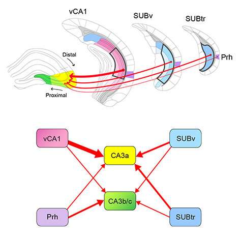
Schematic summary of new hippocampal circuit connections that are revealed in the new PLOS Biology study. The illustration of spatial topology of non-canonical inputs to dorsal hippocampal CA3 (top), and the diagram of differential input strengths and patterns to the dorsal CA3 subregions (bottom) are shown. Credit: UCI School of Medicine

A research team led by University of California, Irvine has discovered new neural circuits that regulate spatial learning and memory in the brain's hippocampal formation. The team identified novel functional roles of new circuit connections between the venal CA1 region and dorsal CA3 regions of the hippocampus and demonstrated that genetic inactivation of this projection impairs object-related spatial learning and memory, but does not modulate anxiety-related behaviors.

29  jan 2022--The study, titled "Non-canonical projections to the hippocampal CA3 regulate spatial learning and memory by modulating the feedforward hippocampal trisynaptic pathway," was published today in PLOS Biology.

The hippocampus is not a homogenous brain area. The septotemporal axis, along which the trisynaptic pathway is located, separates the dorsal region of the hippocampus, which is more involved in learning, memory and spatial navigation, and the ventral region, which plays a role in emotional behavior. The trisynaptic's feedforward, unidirectional circuit organization is well documented, but the connectivity across septal to temporal regions is less well described.

"Our findings extend the knowledge of hippocampal connectivity and its relation to learning and memory processes across the septotemporal axis and provide a circuit foundation to explore these novel functional roles," said Xiangmin Xu, Ph.D., Chancellor's Fellow and professor of anatomy and neurobiology, and director for the Center for Neural Circuit Mapping (CNCM) at the UCI School of Medicine. "The new hippocampal circuit mechanism is highly relevant to treating learning and memory disorders, including Alzheimer's disease."

Building on their earlier work, Xu and his team used multiple viral tracers, including monosynaptic rabies retrograde tracing and herpes (H129)-based anterograde tracing to establish new hippocampal CA1 projections to CA3. Robust mapping results showed that CA1 inputs to CA3 run opposite the trisynaptic pathway and in a temporal to septal direction. They also found that genetic inactivation of the CA1 to CA3 projection impaired object-related spatial learning and memory, but did not modulate anxiety-related behaviors.

"The emergence of viral-genetic mapping techniques enhances our ability to determine the detailed complexity of brain circuity," Xu said. "Our study was made possible by the new viral genetic tools developed by our CNCM investigators at UCI. We are developing these new viral tracers as brain mapping tools, which we plan to share through our center for use by the neuroscience community."


More information: Xiaoxiao Lin et al, Noncanonical projections to the hippocampal CA3 regulate spatial learning and memory by modulating the feedforward hippocampal trisynaptic pathway, PLOS Biology (2021). DOI: 10.1371/journal.pbio.3001127

Yanjun Sun et al, CA1-projecting subiculum neurons facilitate object–place learning, Nature Neuroscience (2019). DOI: 10.1038/s41593-019-0496-y


Why do we forget? New theory proposes 'forgetting' is actually a form of learning

Why do we forget? New theory proposes ‘forgetting’ is actually a form of learning
The new theory proposes that forgetting is due to circuit remodelling that switches engram cells from an accessible to an inaccessible state. Credit: Dr. Nora Raschle

We create countless memories as we live our lives but many of these we forget. Why?

29 jan 2022--Counter to the general assumption that memories simply decay with time, 'forgetting' might not be a bad thing—that is according to scientists who believe it may represent a form of learning.

The scientists behind the new theory—outlined today in the international journal Nature Reviews Neuroscience—suggest that changes in our ability to access specific memories are based on environmental feedback and predictability. Rather than being a bug, forgetting may be a functional feature of the brain, allowing it to interact dynamically with the environment.

In a changing world like the one we and many other organisms live in, forgetting some memories can be beneficial as this can lead to more flexible behavior and better decision-making. If memories were gained in circumstances that are not wholly relevant to the current environment, forgetting them can be a positive change that improves our wellbeing.

So, in effect, the scientists believe we learn to forget some memories while retaining others that are important.

Forgetting of course comes at the cost of lost information, but a growing body of research indicates that, at least in some cases, forgetting is due to altered memory access rather than memory loss.

The new theory has been proposed by Dr. Tomás Ryan, Associate Professor in the School of Biochemistry and Immunology and the Trinity College Institute of Neuroscience at Trinity College Dublin, and Dr. Paul Frankland, Professor in the Department of Psychology at the University of Toronto and the Hospital for Sick Children in Toronto.

Both Dr. Ryan and Dr. Frankland are fellows of the Canadian global research organization CIFAR, which enabled this collaboration through its Child & Brain Development program, which is pursuing interdisciplinary work in this area.

Dr. Ryan, whose research team is based in the Trinity Biomedical Sciences Institute (TBSI), said:

"Memories are stored in ensembles of neurons called 'engram cells' and successful recall of these memories involves the reactivation of these ensembles. The logical extension of this is that forgetting occurs when engram cells cannot be reactivated. The memories themselves are still there, but if the specific ensembles cannot be activated they can't be recalled. It's as if the memories are stored in a safe but you can't remember the code to unlock it.

"Our new theory proposes that forgetting is due to circuit remodeling that switches engram cells from an accessible to an inaccessible state. Because the rate of forgetting is impacted by environmental conditions, we propose that forgetting is actually a form of learning that alters memory accessibility in line with the environment and how predictable it is."

Dr. Frankland added:

"There are multiple ways in which our brains forget, but all of them act to make the engram—the physical embodiment of a memory—harder to access."

Speaking to the case of pathological forgetting in disease, Dr. Ryan and Dr. Frankland note:

"Importantly, we believe that this 'natural forgetting' is reversible in certain circumstances, and that in disease states—such as in people living with Alzheimer's disease for example—these natural forgetting mechanisms are hijacked, which results in greatly reduced engram cell accessibility and pathological memory loss."


More information: Tomás J. Ryan et al, Forgetting as a form of adaptive engram cell plasticity, Nature Reviews Neuroscience (2022). DOI: 10.1038/s41583-021-00548-3

Monday, November 02, 2015

Underlying processes of working memory are more complex than previously thought

hippocampus

The hippocampus is a region of the brain largely responsible for memory formation.


02 nov 2015--In order to retain a piece of information for a short time, working memory is required. The underlying processes are considerably more complex than hitherto assumed, as researchers from the Ruhr-Universität Bochum and Bonn University report in the journal Cell Reports. Two brain states must alternate rhythmically in order for a piece of information to be successfully maintained.
Working memory: maintaining new information for a short time
When we want to remember a new piece of information for a short time, for example a phone number, working memory is called upon. Different brain regions are involved in this process, including the hippocampus, which is known for its crucial role in long-term memory. The team headed by Prof Dr Nikolai Axmacher from the Institute of Cognitive Neuroscience in Bochum and Marcin Leszczynski, researcher in Bochum and at the Department of Epileptology at Bonn University, studied rhythmic activity patterns in the hippocampus while the subjects memorised sequences of numbers or faces.
Two activity states at semi-second intervals
To this end, the team worked with epilepsy patients who had electrodes implanted into the hippocampus for the purpose of surgical planning. Those electrodes enabled the researchers to measure the activity of the region embedded deeply in the brain. While the patients memorised sequences of faces or numbers, the researchers observed two activity states in the hippocampus, which alternated twice per second: an excited and a less excited state.
Seemingly simple tasks require highly complex processes
If the rhythmic pattern did not occur in the hippocampus, the patients tended to make mistakes during the task. Based on the activity patterns, the researchers were also able to estimate how many numbers or faces the test subjects could reliably memorise. "The results show that the brain performs highly complex processes even during seemingly simple tasks," says Prof Nikolai Axmacher. "Our subjective feeling if something is simple or complex is not a reliable marker for how the brain actually solves a task."
More information: M. Leszczyński, J. Fell, N. Axmacher (2015): Rhythmic working memory activation in the human hippocampus, Cell Reports, DOI: 10.1016/j.celrep.2015.09.081 
Provided by Ruhr-Universitaet-Bochum

Saturday, March 24, 2012

Learning best when you rest: Sleeping after processing new info most effective, new study shows

Nodding off in class may not be such a bad idea after all. New research from the University of Notre Dame shows that going to sleep shortly after learning new material is most beneficial for recall.

24 march 2012--Titled "Memory for Semantically Related and Unrelated Declarative Information: The Benefit of Sleep, the Cost of Wake," the study was published March 22 in PLOS One.

Notre Dame Psychologist Jessica Payne and colleagues studied 207 students who habitually slept for at least six hours per night. Participants were randomly assigned to study declarative, semantically related or unrelated word pairs at 9:00 a.m. or 9:00 p.m., and returned for testing 30 minutes, 12 hours or 24 hours later. Declarative memory refers to the ability to consciously remember facts and events, and can be broken down into episodic memory (memory for events) and semantic memory (memory for facts about the world). People routinely use both types of memory every day – recalling where we parked today or learning how a colleague prefers to be addressed.

At the 12-hour retest, memory overall was superior following a night of sleep compared to a day of wakefulness. However, this performance difference was a result of a pronounced deterioration in memory for unrelated word pairs; there was no sleep-wake difference for related word pairs. At the 24-hour retest, with all subjects having received both a full night of sleep and a full day of wakefulness, subjects' memories were superior when sleep occurred shortly after learning, rather than following a full day of wakefulness.

"Our study confirms that sleeping directly after learning something new is beneficial for memory. What's novel about this study is that we tried to shine light on sleep's influence on both types of declarative memory by studying semantically unrelated and related word pairs," Payne says.

"Since we found that sleeping soon after learning benefited both types of memory, this means that it would be a good thing to rehearse any information you need to remember just prior to going to bed. In some sense, you may be 'telling' the sleeping brain what to consolidate."

Provided by University of Notre Dame

Tuesday, July 07, 2009

Can Video Games Boost Thinking Skills In Elderly?

07 july 2009--Researchers at North Carolina State University and the Georgia Institute of Technology have received a $1.2 million grant from the National Science Foundation to study whether and how video games can boost memory and thinking skills in the elderly - and then to use their findings to develop a prototype video game to do just that.

The researchers will use the grant money, which is part of the federal stimulus package, to fund a two-phase research initiative. In phase one, researchers will ascertain whether certain qualities that can be found in video games result in improved cognitive functioning in older adults. Cognitive functioning refers to memory, problem-solving, critical thinking and other mental skills.

Dr. Anne McLaughlin, assistant professor of psychology at NC State and the principal investigator (PI) for the grant, explains that the first phase asks: "What qualities does a game need to contain to improve cognition? We want to determine the components an effective game should have."

In phase two, once the researchers have determined which qualities result in the most significant improvements, they will develop a set of guidelines that can be used to design a new class of video game for older adults, as well as a prototype video game that follows those guidelines. The NSF grant spans four years, from September 2009 to August 2013.

McLaughlin says previous research suggests there are three qualities in video games that may foster improved cognitive functioning: attentional demand, novelty and social interaction. Attentional demand is the degree to which an individual has to focus attention on a task in order to complete it successfully. Novelty, or exposure to a task one has not encountered before, "may also be relevant," McLaughlin says, "because existing research shows that novelty is a catalyst for learning." Social interaction should also encourage players to devote more attention and effort to the game.

The researchers will test the cognitive functioning of participants, men and women 65 years old and older, to establish a baseline. They will then have participants play BOOM BLOX™ or BOOM BLOX™ Bash Party, Wii™ video games developed by Electronic Arts, before testing the participants' cognitive functioning again. BOOM BLOX and BOOM BLOX Bash Party are games in which novelty, attentional demand and social interaction may be manipulated by the researchers.

"For example, if we find that novelty and attentional demand improve cognition, we'll then develop a game that focuses on that," McLaughlin says. Dr. Jason Allaire, an assistant professor of psychology at NC State who is co-PI on the grant, explains that developing a new game highlighting the qualities that seemed to have the greatest impact on cognitive functioning "will allow us to see if we can get greater benefits that might transfer to real-world outcomes such as remembering to take medication."

McLaughlin and Allaire will be working with Georgia Tech Research Scientist Maribeth Gandy to develop the prototype game based on their research findings.

Source:
Matt Shipman
North Carolina State University

Saturday, April 25, 2009

Think memory worsens with age? Then yours probably will

25 april 2009--Thinking your memory will get worse as you get older may actually be a self-fulfilling prophecy. Researchers at North Carolina State University have found that senior citizens who think older people should perform poorly on tests of memory actually score much worse than seniors who do not buy in to negative stereotypes about aging and memory loss.

In a study published earlier this month, psychology professor Dr. Tom Hess and a team of researchers from NC State show that older adults' ability to remember suffers when negative stereotypes are "activated" in a given situation. "For example, older adults will perform more poorly on a memory test if they are told that older folks do poorly on that particular type of memory test," Hess says. Memory also suffers if senior citizens believe they are being "stigmatized," meaning that others are looking down on them because of their age.

"Such situations may be a part of older adults' everyday experience," Hess says, "such as being concerned about what others think of them at work having a negative effect on their performance – and thus potentially reinforcing the negative stereotypes." However, Hess adds, "The positive flip side of this is that those who do not feel stigmatized, or those in situations where more positive views of aging are activated, exhibit significantly higher levels of memory performance." In other words, if you are confident that aging will not ravage your memory, you are more likely to perform well on memory-related tasks.

The study also found a couple of factors that influenced the extent to which negative stereotypes influence older adults. For example, the researchers found that adults between the ages of 60 and 70 suffered more when these negative stereotypes were activated than seniors who were between the ages of 71 and 82. However, the 71-82 age group performed worse when they felt stigmatized.

Finally, the study found that negative effects were strongest for those older adults with the highest levels of education. "We interpret this as being consistent with the idea that those who value their ability to remember things most are the most likely to be sensitive to the negative implications of stereotypes, and thus are most likely to exhibit the problems associated with the stereotype."

"The take-home message," Hess says, "is that social factors may have a negative effect on older adults' memory performance."

Hess is the lead author on the study, "Moderators of and Mechanisms Underlying Stereotype Threat Effects on Older Adults' Memory Performance." Co-authors on the study are former NC State students Joey T. Hinson and Elizabeth A. Hodges. The study was published online April 1 by Experimental Aging Research.

Wednesday, January 28, 2009

Another Potential Benefit of Cutting Calories: Better Memory

By PAM BELLUCK

28 jan 2009--The study, published Monday in The Proceedings of the National Academy of Sciences, appears to be the first to link calorie-restricted diets with improved memory in people. Studies with animals have shown memory improvement, but there is debate about the impact of calorie restriction on humans’ cognitive function.

The study was small, involving 50 men and women ages 50 to 72 who ranged from normal weight to overweight.

Members of one group ate food they normally ate but were instructed to cut their calories by 30 percent, primarily by eating smaller portions, said Dr. Agnes Flöel of the University of Münster in Germany, a neurologist and one of the researchers. Members of a second group kept their calories the same but were instructed to increase the unsaturated fat (healthy fat) they ate by 20 percent. A third group made no dietary changes.

Participants were advised by dietitians but monitored their own eating over three months, Dr. Flöel said. Then they took tests involving memorizing words. The calorie-restricted group averaged 20 percent improvement in memory performance. The other groups showed no significant change.

Dr. Flöel said the memory improvement might be linked to a decrease in insulin and inflammation in the calorie-restricted participants, who lost four to seven pounds.

She said lower insulin levels might “increase the sensitivity of receptors” in the brain and improve insulin signaling, allowing memories to be maintained longer. She said inflammation was believed to “promote aggregation of toxic proteins and promote insulin resistance,” so decreased inflammation would help brain function.

Other scientists said the results were intriguing.

“This is the first that I know of in humans that is showing that effect,” said Grant Brinkworth, a research scientist at the Commonwealth Scientific and Industrial Research Organization in Australia. “The fact that they saw these correlations and quite strong correlations between memory and insulin, and also inflammation markers, suggests that there may be some physiological underpinning to the effect.”

Calorie restriction is being studied intently by researchers. Animal studies have shown that eating less leads to less disease and longer life, but human studies have been mixed on that question.

Its effect on cognitive function is unclear. Some studies have associated self-policed dieting with cognitive decline, but some experts say those dieters might have been preoccupied with thoughts of food and weight loss.

Other research, including part of a federally financed study of calorie restriction over two years, called Calerie, found no decline in cognitive performance, but no improvement either.

A principal investigator on the Calerie study, Eric Ravussin, a professor of human physiology at the Pennington Biomedical Research Center in Baton Rouge, La., said Calerie’s results did not undercut the new study. Calerie “didn’t have the same hard testing” on cognitive function, he said, it did not test memory, and “our subjects were much younger,” ages 20 to 50.

Dr. Flöel said researchers were surprised that participants in the unsaturated fat group showed no memory improvement, but that might have occurred because most did not get their unsaturated fats from fish, which is considered beneficial because it is high in omega-3 fatty acids.

She said her team was conducting a larger study in which the unsaturated fat group is eating a lot of omega-3 fats, and was also planning to study calorie cutting and omega-3 in elderly people with mild cognitive impairment, a precursor to dementia.

Monday, December 22, 2008

Imaging Reveals Age-Related Memory Differences

In older adults, enhanced amygdala activity linked to memory of negative pictures was preserved

22 dec 2008-- Activity in the amygdala linked to subsequent memory of negative images was preserved in older adults, but older individuals had less subsequent-memory activity for negative pictures in visual cortices, according to research published in the January issue of Psychological Science.

Peggy L. St. Jacques, of Duke University in Durham, N.C., and colleagues analyzed functional MRI data from 15 young women (mean age 24.8 years) and 15 older women (mean age 70.2 years). The subjects viewed positive, negative and neutral images and later described the pictures as best as possible after a brief description.

Both age groups used the amygdala more for subsequent memory of negative pictures than neutral ones, the investigators found. Also, brain areas with more subsequent-memory activity for negative than neutral pictures showed a posterior-anterior shift in aging pattern. The results also showed functional connections between the amygdala and the bilateral dorsolateral prefrontal cortices, which is a region involved in emotional regulation, in older subjects, the researchers report.

"Compared with young adults, older adults had greater functional connectivity between the right amygdala and bilateral dorsolateral prefrontal cortices, a possible reflection of increased emotional regulation of negative pictures, but decreased functional connectivity between the amygdala and typical subsequent-memory regions such as the hippocampus, a possible reflection of decreased modulation by the amygdala and decreased memory retrieval for negative pictures. These findings advance understanding of the age-related alterations in the neural networks underlying subsequent memory for negative stimuli when affective function is preserved," the authors write.

Full Text

Friday, November 28, 2008

Brain scans show root of memory glitch with aging

By MALCOLM RITTER
NEW YORK, 28nov 2008 – Brain scans of older people in a noisy lab machine give biological backing to the idea that distraction hampers memory with aging, researchers reported Wednesday.
The finding bolsters a theory about one reason why memory weakens with age: older people have more trouble remembering some things because they're more easily distracted when they try to learn them.
The memory exercise reported in the latest issue of the Journal of Neuroscience dealt with recognizing faces, but the findings apply to the more general task of trying to remember something a person sees or hears, said lead author Dale Stevens.
Stevens, a postdoctoral researcher at Harvard University, did the work while at the Rotman Research Institute at Baycrest, which is affiliated with the University of Toronto.
Older people who have to learn something should do all they can to focus on that task and eliminate potential distractions, he advised.
The study compared 10 healthy people in their 60s and 70s to a dozen younger volunteers, ages 22 to 36. Their brains were scanned while they looked at photographs of people they did not know. As each photograph was displayed for one second, the volunteers were asked if they'd seen it before in the study.
In all they saw 180 different faces, of which 120 showed up a second time. The older participants failed to recognize a face they'd already seen 43 percent of the time, compared to 26 percent for the younger volunteers.
Researchers went back to see what was going on in the brains of the volunteers when they first saw a face that they later failed to recognize. Why didn't those faces get planted in memory?
In both groups, a brain area called the hippocampus, which is involved in memory, was less active when a face failed to stick in the memory than when it did. That was no surprise. More interestingly, the older group also showed heightened activity in certain other brain areas while the younger volunteers did not.
Those areas included the auditory cortex, which plays a role in analyzing sound, and several areas involved in directing attention, Stevens said.
So what was going on? The brain-scanning machine was noisy, with lots of knocking, buzzing and banging like a jackhammer, Stevens said. Even with the earplugs the volunteers wore, "it's a little distracting," he said.
The brain activity in the older volunteers shows that the noise was more distracting to them than to the younger participants, and reveals the brain circuits involved, researchers concluded.
The study could not address when a person's brain starts to act up this way. But Cheryl Grady of the Rotman institute, another author, said she suspects it may begin between ages 40 and 60.
Dr. Barry Gordon, a neurology professor and memory expert at The Johns Hopkins Medical Institutions in Baltimore, called the work "an appreciable advance." A next step could be seeing whether older people will do better on a memory test if they're warned about the distraction problem, said Gordon, who wasn't involved in the new study.
In any case, he said, "if you want to remember something, it's more important if you're older than younger not to be listening to your iPod."
___
On the Net:
The Journal of Neuroscience: http://www.jneurosci.org/

Saturday, September 06, 2008


For the Brain, Remembering Is Like Reliving


By BENEDICT CAREY
06 sept 2008--Scientists have for the first time recorded individual brain cells in the act of summoning a spontaneous memory, revealing not only where a remembered experience is registered but also, in part, how the brain is able to recreate it.
The recordings, taken from the brains of epilepsy patients being prepared for surgery, demonstrate that these spontaneous memories reside in some of the same neurons that fired most furiously when the recalled event had been experienced. Researchers had long theorized as much but until now had only indirect evidence.
Experts said the study had all but closed the case: For the brain, remembering is a lot like doing (at least in the short term, as the research says nothing about more distant memories).
The experiment, being reported Friday in the journal Science, is likely to open a new avenue in the investigation of Alzheimer’s disease and other forms of dementia, some experts said, as well as help explain how some memories seemingly come out of nowhere. The researchers were even able to identify specific memories in subjects a second or two before the people themselves reported having them.
“This is what I would call a foundational finding,” said Michael J. Kahana, a professor of psychology at the University of Pennsylvania, who was not involved in the research. “I cannot think of any recent study that’s comparable.
“It’s a really central piece of the memory puzzle and an important step in helping us fill in the detail of what exactly is happening when the brain performs this mental time travel” of summoning past experiences.
The new study moved beyond most previous memory research in that it focused not on recognition or recollection of specific symbols but on free recall — whatever popped into people’s heads when, in this case, they were asked to remember short film clips they had just seen.
This ability to richly reconstitute past experience often quickly deteriorates in people with Alzheimer’s and other forms of dementia, and it is fundamental to so-called episodic memory — the catalog of vignettes that together form our remembered past.
In the study, a team of American and Israeli researchers threaded tiny electrodes into the brains of 13 people with severe epilepsy. The electrode implants are standard procedure in such cases, allowing doctors to pinpoint the location of the mini-storms of brain activity that cause epileptic seizures.
The patients watched a series of 5- to 10-second film clips, some from popular television shows like “Seinfeld” and others depicting animals or landmarks like the Eiffel Tower. The researchers recorded the firing activity of about 100 neurons per person; the recorded neurons were concentrated in and around the hippocampus, a sliver of tissue deep in the brain known to be critical to forming memories.
In each person, the researchers identified single cells that became highly active during some videos and quiet during others. More than half the recorded cells hummed with activity in response to at least one film clip; many of them also responded weakly to others.
After briefly distracting the patients, the researchers then asked them to think about the clips for a minute and to report “what comes to mind.” The patients remembered almost all of the clips. And when they recalled a specific one — say, a clip of Homer Simpson — the same cells that had been active during the Homer clip reignited. In fact, the cells became active a second or two before people were conscious of the memory, which signaled to researchers the memory to come.
“It’s astounding to see this in a single trial; the phenomenon is strong, and we were listening in the right place,” said the senior author, Dr. Itzhak Fried, a professor of neurosurgery at the University of California, Los Angeles, and the University of Tel Aviv.
His co-authors were Hagar Gelbard-Sagiv, Michal Harel and Rafael Malach of the Weizmann Institute of Science in Israel, and Roy Mukamel, of U.C.L.A.
Dr. Fried said in a phone interview that the single neurons recorded firing most furiously during the film clips were not acting on their own; they were, like all such cells, part of a circuit responding to the videos, including thousands, perhaps millions, of other cells.
In studies of rodents, including a paper that will also appear Friday in the journal Science, neuroscientists have shown that special cells in the hippocampus are sensitive to location, activating when the animal passes a certain spot in a maze. The firing pattern of these cells forms the animals’ spatial memory and can predict which way the animal will turn, even if it makes a wrong move.
Some scientists argue that as humans evolved, these same cells adapted to register a longer list of elements — including possibly sounds, smells, time of day and chronology — when an experience occurred in relation to others.
Single-cell recordings cannot capture the entire array of circuitry involved in memory, which may be widely distributed beyond the hippocampus area, experts said. And as time passes, memories are consolidated, submerged, perhaps retooled and often entirely reshaped when retrieved later.
Though it did not address this longer-term process, the new study suggests that at least some of the neurons that fire when a distant memory comes to mind are those that were most active back when it happened, however long ago that was.
“The exciting thing about this,” said Dr. Kahana, the University of Pennsylvania professor, “is that it gives us direct biological evidence of what before was almost entirely theoretical.”

Friday, August 17, 2007

'Memory molecule' stores memories in neocortex

PKMzeta required for memory to endure
The “memory storage molecule” – PKMzeta – maintains long-term memories in the neocortex and its presence is continually required in order for the memory to endure, according to a finding by researchers at the Weizmann Institute in Rehovot, Israel and SUNY Downstate Medical Center in Brooklyn, New York. The study results were published in the current edition of Science magazine (www.sciencemag.org). The title of the paper is “Rapid Erasure of Long-Term Memory Associations in Cortex by an Inhibitor of PKMzeta.”
It was previously determined that PKMzeta in the hippocampus – an area of the brain involved in navigation and the initial storage of memory – was necessary to preserve spatial memory; but little was known about PKMzeta activity in the neocortex, the part of the brain thought to be responsible for permanently storing most long-term memories, including those required for higher-level cognitive functions, such as language and complex thought.
This new finding – that inhibiting PKMzeta causes the rapid loss of neocortical memories learned even weeks before – means that persistent phosphorylation by PKMzeta in the neocortex is necessary to store these long-term memories and has potential clinical significance, for example, in the field of cognitive enhancement.

Tuesday, June 05, 2007

Forgetting May Be Part of the Process of Remembering

Whether drawing a mental blank on a new A.T.M. password, a favorite recipe or an old boyfriend, people have ample opportunity every day to curse their own forgetfulness. But forgetting is also a blessing, and researchers reported on Sunday that the ability to block certain memories reduces the demands on the brain when it is trying to recall something important.
The study, appearing in the journal Nature Neuroscience, is the first to record visual images of people’s brains as they suppress distracting memories. The more efficiently that study participants were tuning out irrelevant words during a word-memorization test, the sharper the drop in activity in areas of their brains involved in recollection. Accurate remembering became easier, in terms of the energy required.
Blocking out a distracting memory is something like ignoring an old (and perhaps distracting) acquaintance, experts say: it makes it that much harder to reconnect the next time around. But recent studies suggest that the brain plays favorites with memories in exactly this way, snubbing some to better capture others. A lightning memory, in short, is not so much a matter of capacity as it is of ruthless pruning — and the new study catches the trace of this process at it happens.
“We’ve argued for some time that forgetting is adaptive, that people actively inhibit some memories to facilitate mental focus,” as when they are trying to recall a friend’s new phone number or the location of a parking space, said Michael Anderson, a professor of cognitive neuroscience at the University of Oregon.
Dr. Anderson, who was not involved in the new research, said it was “ important new work because it maps out how this is happening neurobiologically.”
The researchers, neuroscientists at Stanford University, used a memory test intended to gauge how well people can recall studied words from among many similar words they have also seen. They had 20 young men and women, mostly Stanford students, view in quick succession a list of 240 word pairs. These included 40 capitalized words, each paired with six related, lower-case words: For example, “ATTIC-dust,” “ATTIC-junk,” and so on.
After studying the pairs, the participants were instructed to memorize three selected pairs from each of 20 capitalized words. In effect, this forced them to flag individual pairs, like ATTIC-dust, while trying to tune out very similar, distracting ones, like ATTIC-junk, for half of the total list of pairs they saw. They were told not to memorize any pairs from the other half of the list.
The researchers tested each person’s memory several times, and found that scores ranged from about 30 percent accuracy to 80 percent. They also measured how well each person suppressed the distracting word pairs, by comparing recall of those pairs with recall of the half of the list that was studied at first but later ignored. All the testing was done while participants were having their brains scanned by an M.R.I. machine.
“We found that the magnitude of the decrease in activity on M.R.I. was correlated to the amount of weakening of these competing memories” when the subjects were recalling the target words, said Brice Kuhl, a graduate student in the psychology department at Stanford and the study’s lead author. His co-authors were Anthony Wagner, Nicole Dudukovic and Itamar Kahn.
In particular, the researchers found that the more a study participant had suppressed the memory of distracting word pairs, the steeper the decrease in activity in a region of the brain called the anterior cingulated cortex. This neural area is especially active when people are engaged in weighing choices, say, in choosing which card to play in a game of hearts with two or more good options.
“From a broader point of view, given what we know about this area, the activity decreases as the task becomes more automatic, less demanding,” said Dr. Wagner, the senior author.
People blank on new passwords so often because of the distracting presence of old or other current passwords. The better the brain can block those distracting digits, the easier it can bring to mind the new ones, Dr. Wagner said.
This process is extremely familiar to people who have been immersed in a foreign language. In a recent study of native English speakers led by Dr. Anderson, researchers showed that beginners being drilled in Spanish were very slow to link pictures and words in English, compared with more bilingual participants. Those fluent in both languages had resolved the competition between the two tongues, inhibiting the encroachment, for example, of the word “zapato” on the word “shoe.”
In all, this research suggests that memories are more often crowded out than lost. An ideal memory improvement program, Dr. Anderson said, “would include a course on how to impair your memory. Your head is full of a surprising number of things that you don’t need to know.”
The findings should also reduce some of the anxiety surrounding “senior moments,” researchers say. Some names, numbers and details are hard to retrieve not because memory is faltering, but because it is functioning just as it should.

Wednesday, April 18, 2007

Sleep enforces the temporal sequence in memory

We have usually quite strong memories of past events like an exciting holiday or a jolly birthday party. However it is not clear how the brain keeps track of the temporal sequence in such memories: did Paul spill a glass of wine before or after Mary left the party?
Previous findings from a research group headed by Jan Born at the University of Lübeck have confirmed the widely held view that long-term memories are formed particularly during sleep, and that this process relies on the brain replaying recently encoded experiences during the night. The same research group now provides evidence that sleep not only strengthens the content of a memory but also the particular order in which they were experienced, probably by a replay of the experiences in "forward" direction.
Students were asked to learn triplets of words presented one after the other. Afterwards they slept, whereas in a control condition no sleep was allowed. Later, recall was tested by presenting one word and asking which one came before and which one came after during learning. Sleep was found to enhance word recall, but only when the students were asked to reproduce the learned words in forward direction.
This finding shows that sleep associated consolidation of memories enforces the temporal structure of the memorized episode that otherwise might be blurred to a timeless puzzle of experiences.

Tuesday, March 20, 2007

New Reason To Hit The Gym: Fighting Memory Loss

Research has shown that people who exercise do better on memory tests. Now a new Columbia University Medical Center study explains specifically what exercise does within the brain. Exercise, the researchers found, targets a region of the brain within the hippocampus, known as the dentate gyrus, which underlies normal age-related memory decline that begins around age 30 for most adults.
This finding is significant because it was accomplished via the first-ever observation of neurogenesis, the growth of neurons, within a living brain. Using an MRI imaging technique developed at Columbia, the researchers were able to identify neurogenesis within the dentate gyrus region following exercise. Previously, researchers were only able to prove neurogenesis upon postmortem exam in animal studies.
http://www.sciencedaily.com/releases/2007/03/070320073516.htm

Friday, March 09, 2007

Want a better memory? Stop and smell the roses

People who want to learn things might do better by simply stopping to smell the roses, researchers reported on Thursday.
German researchers found they could use odors to re-activate new memories in the brains of people while they slept -- and the volunteers remembered better later.
Writing in the journal Science, they said their study showed that memories are indeed consolidated during sleep, and show that smells and perhaps other stimuli can reinforce brain learning pathways.
Jan Born of the University of Lubeck in Germany and colleagues had 74 volunteers learn to play games similar to the game of "Concentration" in which they must find matched pairs of objects or cards by turning only one over at a time.
While doing this task, some of the volunteers inhaled the scent of roses. The volunteers then agreed to sleep inside an MRI tube. Functional magnetic resonance imaging was used to "watch" their brains while they slept.

http://news.yahoo.com/s/nm/20070309/hl_nm/memory_smells_dc