Wednesday, March 09, 2022


Damage early in Alzheimer's disease identified via novel MRI approach
Damage early in Alzheimer’s disease ID’d via novel MRI approach
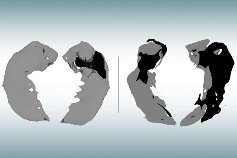
An MRI scan of the hippocampus, the brain’s memory center, in an older person with no signs of cognitive decline (left) and a person of similar age with mild Alzheimer’s (right) has been analyzed with a new technique that shows where healthy brain cells have been lost (dark areas). New research from Washington University School of Medicine in St. Louis shows that this novel MRI approach can identify brain cell damage in people at early stages of Alzheimer’s, before tissue shrinkage is visible on traditional MRI scans. Credit: Satya Kothapalli/School of Medicine

Alzheimer's disease usually is diagnosed based on symptoms, such as when a person shows signs of memory loss and difficulty thinking. Up until now, MRI brain scans haven't proven useful for early diagnosis in clinical practice. Such scans can reveal signs of brain shrinkage due to Alzheimer's, but the signs only become unmistakable late in the course of the disease, long after the brain is significantly damaged and most people have been diagnosed via other means.

09 mar 2022--But new research from Washington University School of Medicine in St. Louis shows that a mathematical analysis of data obtained with a novel MRI approach can identify brain cell damage in people at early stages of Alzheimer's, before tissue shrinkage is visible on traditional MRI scans and before cognitive symptoms arise.

"This could be a new way to use MRI to diagnose people with Alzheimer's before they develop symptoms," said senior author Dmitriy Yablonskiy, Ph.D., a professor of radiology at the university's Mallinckrodt Institute of Radiology. "The technique takes only six minutes to acquire data and can be implemented on MRI scanners that are already used worldwide for patient diagnostics and clinical trials."

Published in the Journal of Alzheimer's Disease, the study relies on a new quantitative Gradient Echo (qGRE) MRI technique developed in the Yablonskiy lab to show brain areas that are no longer functioning due to a loss of healthy neurons.

"Using this technique in patients with Alzheimer's disease, we discovered brain areas that look normal on traditional MRI but look dark on qGRE images, which we attribute to significant neurodegeneration," said Satya V. V. N. Kothapalli, Ph.D., a staff scientist in radiology and the first author of the study. "We call them 'dark matter.'"

While traditional MRI is capable of showing where damaged areas of the brain have decreased in volume, the qGRE technique goes a step further, detecting the loss of neurons that precedes brain shrinkage and cognitive decline.

Alzheimer's disease develops slowly over the course of two decades or more before symptoms arise. First the brain protein amyloid beta accumulates into plaques in the brain, then another brain protein—tau—coalesces into tangles and neurons begin to die. Finally, tissue atrophy becomes visible on MRI brain scans, and cognitive symptoms arise. People at early stages of the disease can be identified via amyloid-PET brain scans or by testing for amyloid in the blood or the cerebrospinal fluid that surrounds the brain and , but such tests do not provide information on neuronal damage.

The study involved 70 people ages 60 to 90 who were recruited through the Charles F. and Joanne Knight Alzheimer Disease Research Center (Knight ADRC). Participants completed extensive clinical and cognitive testing to assess their level of cognitive impairment. The participant group included people with no cognitive impairment as well as those with very mild, mild or moderate impairments.

Each participant underwent either a PET brain scan or a spinal tap to gauge the amount of amyloid plaques in his or her brain. They also underwent MRI brain scans.

Researchers applied the qGRE MRI technique to scan the hippocampus, the brain's memory center and one of the earliest affected brain regions in Alzheimer's. Results showed the hippocampus often contained a viable tissue section with relatively preserved neurons and a dark matter dead zone virtually devoid of healthy neurons.

These dark matter areas were present in people who tested positive for amyloid but were not yet experiencing symptoms, and they grew larger as the disease progressed. Compared with traditional MRI measures of brain atrophy, biomarkers for dark matter correlated much better with individual cognitive scores for very mild to moderate dementia.

The study builds on and corroborates findings from Alzheimer's research that took place at Washington University more than two decades ago when Alzheimer's was formally diagnosed only through autopsy.

In 2001, John C. Morris, MD, the Harvey A. and Dorismae Hacker Friedman Distinguished Professor of Neurology and director of the Knight ADRC, led a study that examined the brain tissue of deceased Alzheimer's patients and found that damaged brain regions had begun to lose healthy neurons well before the disease caused a significant loss of brain volume in these areas.

Then, in the early 2000s, Tammie L. S. Benzinger, MD, Ph.D., a professor of radiology and of neurosurgery, and the Knight ADRC's director of imaging studies, was among the pioneers at the Mallinckrodt Institute of Radiology to use PET brain scans targeted against amyloid beta as a tool for detecting Alzheimer's.

In the current study, also co-authored by Morris and Benzinger, researchers documented the same relationship between neuronal loss and Alzheimer's symptoms using the non-invasive qGRE MRI technique in living patients.

Working with co-author Richard Perrin, MD, Ph.D., an associate professor of pathology and immunology, the research team also confirmed this relationship under the microscope by examining brain tissues that were donated after the death of a study participant. The postmortem examination showed that the neuronal loss in the hippocampus actually exceeds loss of tissue volume and that these changes are well reflected in MRI measures of dark matter.

Yablonskiy and colleagues are among the many researchers now pursuing a low-cost, easily accessible test for Alzheimer's as an alternative to the expensive PET brain scans and invasive spinal taps now used in research settings to assess the presence and progression of the disease.

Such a test, especially one that can identify people at very early stages of disease, would provide a huge boost to Alzheimer's research, drastically slashing the cost and the time necessary to screen patients for clinical trials, thus spurring the development of new treatments.

While Alzheimer's researchers continue to pursue drug treatments for the disease, most agree that successful treatment will hinge on early detection and on finding ways to head off brain damage before later stages of Alzheimer's.

While PET scans and spinal taps continue to play important roles in Alzheimer's research, both have limitations that prevent their widespread use as a screening tool for early signs of the disease.

PET brain scans are still the gold standard for detecting signs of Alzheimer's, but the machines are expensive and seldom available for routine patient diagnosis, much less widespread clinical screening. PET scans also require the injection of a radioactive tracer for brain imaging.

As Alzheimer's progresses, it can be detected by testing for tau proteins in cerebrospinal fluid, but sampling requires a spinal tap that may be too invasive for use as a general screening tool, especially for people who have no symptoms.

Another promising option for Alzheimer's screening—also under development at Washington University—is a noninvasive, relatively inexpensive blood test that has proven to be highly accurate in detecting early signs of Alzheimer's disease. A commercial version of the blood test recently became available to doctors in the United States and Europe, but it is not yet covered by health insurance.

While each testing approach has its own strengths and weaknesses, the qGRE MRI technique may be poised for early adoption since it is based on MRI technology that is widely available worldwide, is noninvasive and can be carried out without the use of radioactive tracers.

"Our qGRE test offers great potential as an early diagnostic tool for the preclinical stage of Alzheimer's disease, thus providing a large window for therapeutic intervention," Yablonskiy said. "It also has great potential as a noninvasive MRI technique available in a conventional clinical setting for the widespread screening that is necessary to get people with early Alzheimer's into clinical drug trials."


More information: Satya V.V.N. Kothapalli et al, Quantitative Gradient Echo MRI Identifies Dark Matter as a New Imaging Biomarker of Neurodegeneration that Precedes Tisssue Atrophy in Early Alzheimer's Disease, Journal of Alzheimer's Disease (2021). DOI: 10.3233/JAD-210503
Journal information: Journal of Alzheimer's Disease 


More alcohol, less brain: Study finds an association that begins with an average of just one drink a day

brain
Credit: Pixabay/CC0 Public Domain

The science on heavy drinking and the brain is clear: The two don't have a healthy relationship. People who drink heavily have alterations in brain structure and size that are associated with cognitive impairments.

09 mar 2022--But according to a new study, alcohol consumption even at levels most would consider modest—a few beers or glasses of wine a week—may also carry risks to the brain. An analysis of data from more than 36,000 adults, led by a team from the University of Pennsylvania, found that light-to-moderate alcohol consumption was associated with reductions in overall brain volume.

The link grew stronger the greater the level of alcohol consumption, the researchers showed. As an example, in 50-year-olds, as average drinking among individuals increases from one alcohol unit (about half a beer) a day to two units (a pint of beer or a glass of wine) there are associated changes in the brain equivalent to aging two years. Going from two to three alcohol units at the same age was like aging three and a half years. The team reported their findings in the journal Nature Communications.

"The fact that we have such a large sample size allows us to find subtle patterns, even between drinking the equivalent of half a beer and one beer a day," says Gideon Nave, a corresponding author on the study and faculty member at Penn's Wharton School. He collaborated with former postdoc and co-corresponding author Remi Daviet, now at the University of Wisconsin-Madison, and Perelman School of Medicine colleagues Reagan Wetherill—also a corresponding author on the study—and Henry Kranzler, as well as other researchers.

"These findings contrast with scientific and governmental guidelines on safe drinking limits," says Kranzler, who directs the Penn Center for Studies of Addiction. "For example, although the National Institute on Alcohol Abuse and Alcoholism recommends that women consume an average of no more than one drink per day, recommended limits for men are twice that, an amount that exceeds the consumption level associated in the study with decreased brain volume,"

Ample research has examined the link between drinking and brain health, with ambiguous results. While strong evidence exists that heavy drinking causes changes in brain structure, including strong reductions in gray and white matter across the brain, other studies have suggested that moderate levels of alcohol consumption may not have an impact, or even that light drinking could benefit the brain in older adults.

These earlier investigations, however, lacked the power of large datasets. Probing massive quantities of data for patterns is the specialty of Nave, Daviet, and colleagues, who have conducted previous studies using the UK Biobank, a dataset with genetic and medical information from half a million British middle-aged and older adults. They employed biomedical data from this resource in the current study, specifically looking at brain MRIs from more than 36,000 adults in the Biobank, which can be used to calculate white and gray matter volume in different regions of the brain.

"Having this dataset is like having a microscope or a telescope with a more powerful lens," Nave says. "You get a better resolution and start seeing patterns and associations you couldn't before."

To gain an understanding of possible connections between drinking and the brain, it was critical to control for confounding variables that could cloud the relationship. The team controlled for age, height, handedness, sex, smoking status, socioeconomic status, genetic ancestry, and county of residence. They also corrected the brain-volume data for overall head size.

The volunteer participants in the Biobank had responded to survey questions about their alcohol consumption levels, from complete abstention to an average of four or more alcohol units a day. When the researchers grouped the participants by average-consumption levels, a small but apparent pattern emerged: The gray and white matter volume that might otherwise be predicted by the individual's other characteristics was reduced.

Going from zero to one alcohol units didn't make much of a difference in brain volume, but going from one to two or two to three units a day was associated with reductions in both gray and white matter.

"It's not linear," says Daviet. "It gets worse the more you drink."

Even removing the heavy drinkers from the analyses, the associations remained. The lower brain volume was not localized to any one brain region, the scientists found.

To give a sense of the impact, the researchers compared the reductions in brain size linked with drinking to those that occur with aging. Based on their modeling, each additional alcohol unit consumed per day was reflected in a greater aging effect in the brain. While going from zero to a daily average of one alcohol unit was associated with the equivalent of a half a year of aging, the difference between zero and four drinks was more than 10 years of aging.

In future work, the authors hope to tap the UK Biobank and other large datasets to help answer additional questions related to alcohol use. "This study looked at average consumption, but we're curious whether drinking one beer a day is better than drinking none during the week and then seven on the weekend," Nave says. "There's some evidence that binge drinking is worse for the brain, but we haven't looked closely at that yet."

They'd also like to be able to more definitively pin down causation rather than correlation, which may be possible with new longitudinal biomedical datasets that are following young people as they age.

"We may be able to look at these effects over time and, along with genetics, tease apart causal relationships," Nave says.

And while the researchers underscore that their study looked only at correlations, they say the findings may prompt drinkers to reconsider how much they imbibe.

"There is some evidence that the effect of drinking on the brain is exponential," says Daviet. "So, one additional drink in a day could have more of an impact than any of the previous drinks that day. That means that cutting back on that final drink of the night might have a big effect in terms of brain aging."

In other words, Nave says, "the people who can benefit the most from drinking less are the people who are already drinking the most."


More information: Associations between alcohol consumption and gray and white matter volumes in the UK Biobank, Nature Communications (2022). DOI: 10.1038/s41467-022-28735-5
Journal information: Nature Communications 


GlyNAC supplementation extends life span in mice

mice
Credit: Pixabay/CC0 Public Domain

"Just like a car is subject to wear and tear over many years of use, limiting how long it works well, the natural aging process can progressively deteriorate the body and limit lifespan," said Rajagopal Sekhar.

09 mar 2022--For over two decades, Sekhar has studied natural aging in older humans and aged mice. His work contributes to providing a better understanding of how glutathione deficiency, increased oxidative stress, mitochondrial dysfunction and multiple additional hallmark defects of aging contribute to the aging process and how they can be reversed with GlyNAC supplementation.

"Energy is the currency of life and is generated by mitochondria. However, aging is associated with mitochondrial malfunction, and this could affect energy availability. When we discovered that GlyNAC supplementation can correct mitochondrial defects in aging, we asked an important question: Could GlyNAC supplementation from a younger age extend length of life?" said Sekhar. "To answer this question, we worked with healthy, normal laboratory mice."

The researchers housed the mice under stable environmental conditions. When mice reach an age of 65-weeks, they typically begin to show a drop in glutathione levels and develop mitochondrial dysfunction and oxidative stress. At this age, Sekhar's team switched the diets of half of the mice to receive GlyNAC, and the other half continued their diet without the supplement. Except for the GlyNAC supplementation, the diets of both groups were the same regarding the content of protein, fat and carbohydrates. Then, the researchers let the mice continue aging undisturbed and recorded how long they lived.

"We were excited to find that the mice that received GlyNAC lived 24% longer than those that did not receive GlyNAC," Sekhar said. "We next wanted to understand how GlyNAC works."

For this, the team conducted a second study in aged mice to investigate mitochondrial dysfunction, glutathione levels, oxidative stress and other hallmarks of aging specifically in the animals' heart, liver and kidneys, and determined the effect of supplementing GlyNAC on these cellular defects. The researchers chose these organs because they conduct vital functions in the body.

In aged mice, the team found that all three organs had glutathione deficiency, oxidative stress, mitochondrial dysfunction, abnormal mitophagy (difficulty in disposing damaged mitochondria), impaired nutrient sensing and genomic damage. GlyNAC supplementation improved and corrected all these defects in the old mice. The researchers propose that it is the improvement of these fundamental biological defects that contributes to the animals' longer life.

"But mice are not humans. Could this happen in people? There is published evidence from our human studies showing that GlyNAC supplementation improves similar defects in people," Sekhar said.

For instance, in a pilot human clinical trial in older adults, Sekhar's group showed that taking GlyNAC for 24-weeks improved many characteristic defects of aging, including glutathione deficiency, oxidative stress, mitochondrial dysfunction, inflammation, insulin resistance, endothelial dysfunction, body fat and genomic damage. Importantly, the researchers also observed improvement in muscle strength, gait speed, exercise capacity and cognitive function.

Another exploratory human trial by his team investigated premature aging in people with HIV and showed that taking GlyNAC improved and corrected multiple similar defects to reverse premature aging and also increased strength, energy levels, cognitive function and quality of life.

Sekhar thinks that the glycine and NAC (from GlyNAC), combined with glutathione (that GlyNAC helps generate), provide the benefits described above and he calls this combination the 'power of 3.'

"There is growing interest in being able to improve both healthy aging and longer life, but this is not an easy task. We have investigated aging in  and in human studies for two decades, and our studies show that GlyNAC supplementation successfully and consistently improves many age-related defects," Sekhar said. "It is exciting that something as simple as GlyNAC can improve several important defects in aging and also extend life."

The research was published in Nutrients.


More information: Premranjan Kumar et al, GlyNAC (Glycine and N-Acetylcysteine) Supplementation in Mice Increases Length of Life by Correcting Glutathione Deficiency, Oxidative Stress, Mitochondrial Dysfunction, Abnormalities in Mitophagy and Nutrient Sensing, and Genomic Damage, Nutrients (2022). doi.org/10.3390/nu14051114

Baylor College of Medicine holds an issued patent on the unique combination of Glycine and NAC (GlyNAC) and has licensed it to Nestlé Health Science for use in Celltrient™ Protect and other products. Nestlé Health Science did not provide any funding or support for this research and was not involved with any aspect of this research. Internal funding from Baylor College of Medicine supported this study. Premranjan Kumar and Ob W. Osahon are members of Sekhar's lab called the Translational Metabolism Unit at Baylor College of Medicine and contributed to this work.


Study: Some of the world's lowest rates of dementia found in Amazonian indigenous groups

amazon
Credit: CC0 Public Domain

As scientists around the world seek for solutions for Alzheimer's disease, a new study reveals that two indigenous groups in the Bolivian Amazon have among the lowest rates of dementia in the world.

09 mar 2022--An international team of researchers found among older Tsimane and Moseten people, only about 1% suffer from dementia. In contrast, 11% of people age 65 and older living in the United States have dementia, according to the Alzheimer's Association.

"Something about the pre-industrial subsistence lifestyle appears to protect older Tsimane and Moseten from dementia," said Margaret Gatz, the lead study author and professor of psychology, gerontology and preventive medicine at the Center for Economic and Social Research at the USC Dornsife College of Letters, Arts and Sciences.

Researchers used computed tomography (CT) brain scan images, cognitive and neurological assessments and culturally appropriate questionnaires—facilitated by a local team of trained translators and Bolivian physicians—to diagnose dementia and cognitive impairment among the Tsimane and Moseten.

The study, published in Alzheimer's & Dementia: The Journal of the Alzheimer's Association, found only five cases of dementia among 435 Tsimane people and just one case among 169 Moseten age 60 and over.

In the same over-60 groups, the research team diagnosed about 8% of Tsimane and 10% of Moseten with mild cognitive impairment (MCI), which is typically marked by early stage memory loss or decline of other cognitive ability, such as language or spatial perception. The study's authors pointed out these rates are more comparable to MCI in high-income countries like the U.S.

Researchers were surprised to find that the study participants found to have dementia or MCI frequently had unusual and prominent calcifications of their intracranial arteries. These study participants frequently displayed parkinsonian symptoms during neurological examinations and cognitive deficits in attention, spatial awareness and executive functioning.

Although calcifications were more common among the cognitively impaired, researchers also observed these vascular calcifications in the CT scans of those without dementia or MCI. They say more research is needed to understand the role of vascular factors as well as infectious and inflammatory disorders—which are highly prevalent in these populations—along with other risks for dementia. To this end, the research team is currently returning to all the Tsimane and Moseten villages to revisit those who were previously assessed.

The roughly 17,000 Tsimane remain physically very active throughout their lifespans as they fish, hunt and farm with hand tools and gather food from the forest. The 3,000 Moseten also reside in rural villages and engage in subsistence agricultural work. Unlike the more isolated Tsimane, they live closer to towns and have schools, access to clean water and medical services, and are more likely to be literate.

The study authors compared their results to a systematic review of 15 studies of indigenous populations in Australia, North America, Guam and Brazil. That earlier review found dementia prevalence ranging from 0.5% to 20% among indigenous older adults.

The fact that indigenous populations in other parts of the world have high rates of dementia may be due to a higher amount of contact with—and adoption of lifestyles of—their non-indigenous neighbors. They also face greater risks of diabetes, hypertension, alcohol abuse, obesity and cardiovascular disease.

These dementia risk factors are extremely low among the Tsimane and Moseten populations. Prior research published in The Lancet showed the Tsimane people have extraordinarily healthy hearts in older age and the lowest prevalence of coronary atherosclerosis (a disease that shows in the form of fatty deposits inside arteries) of any population known to science. This distinction may be linked to their subsistence lifestyle.

Another study published last year in The Journal of Gerontology—led by USC assistant professor Andrei Irimia, also a co-author on the new publication—found that the Tsimane experience less brain atrophy than their American and European peers.

Researchers say in contrast to the Tsimane, lifestyle factors in higher-income countries—including lack of physical activity and diets rich in sugars and fats—contribute to heart disease and may also accelerate brain aging.

A race for Alzheimer's disease solutions

Aging is the most important known risk factor for Alzheimer's and other dementias. Converging evidence points to low formal education, midlife hypertension and diabetes, cardiovascular diseases, physical inactivity and—most recently—air pollution as the major modifiable risk factors for dementia and Alzheimer's disease.

An aging global population, along with the proliferation of those modifiable risk factors, will lead to a tripling of the number of people with dementia worldwide by 2050, to more than 152 million, according to estimates.

"We're in a race for solutions to the growing prevalence of Alzheimer's disease and related dementias," said Hillard Kaplan, a study co-author and professor of health economics and anthropology at Chapman University who has studied the Tsimane for two decades. "Looking at these diverse populations augments and accelerates our understanding of these diseases and generate new insights."

"By working with populations like the Tsimane and the Moseten, we can get a better understanding of global human variation and what human health was like in different environments before industrialization," said Benjamin Trumble, a study co-author and an associate professor in the School of Human Evolution and Social Change and the Center for Evolution and Medicine at Arizona State University. "What we do know is the sedentary, urban, industrial life is quite novel when compared with how our ancestors lived for more than 99% of humanity's existence."

More information: Margaret Gatz et al, Prevalence of dementia and mild cognitive impairment in indigenous Bolivian forager‐horticulturalists, Alzheimer's & Dementia (2022). DOI: 10.1002/alz.12626
Journal information: The Lancet 

Sunday, February 20, 2022


Social isolation and loneliness increase heart disease risk in senior women

Social isolation and loneliness increase heart disease risk in senior women
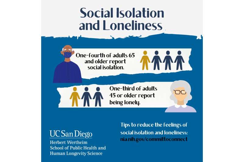
As social networks shrink, older adults are more at risk for social isolation and loneliness. Credit: Herbert Wertheim School of Public Health and Human Longevity Science at University of California San Diego

During the current pandemic, social distancing has been one tool used to reduce the spread of COVID-19. But data from a new study point to as much as a 27% increase in heart disease risk in postmenopausal women who experience both high levels of social isolation and loneliness.

20 feb 2022--The findings of the prospective study, published in the February 2, 2022 online issue of JAMA Network Open, reveal that social isolation and loneliness independently increased cardiovascular disease risk by 8% and 5% respectively. If women experienced high levels of both, their risk rose 13% to 27% compared to women who reported low levels of social isolation and low levels of loneliness.

"We are social beings. In this time of COVID-19, many people are experiencing social isolation and loneliness, which may spiral into chronic states," said first author Natalie Golaszewski, Ph.D., a postdoctoral scholar at the Herbert Wertheim School of Public Health and Human Longevity Science at University of California San Diego. "It is important to further understand the acute and long-term effects these experiences have on cardiovascular health and overall well-being."

Importantly, social isolation and loneliness are mildly correlated and can occur at the same time, but they are not mutually exclusive. A socially isolated person is not always lonely and conversely a person experiencing loneliness is not necessarily socially isolated.

"Social isolation is about physically being away from people, like not touching or seeing or talking to other people. Loneliness is a feeling, one that can be experienced even by people who are regularly in contact with others," said senior author John Bellettiere, Ph.D., M.P.H., assistant professor of epidemiology at the Herbert Wertheim School of Public Health.

Social isolation and loneliness are a growing public health concern as they are associated with health conditions that increase the risk of cardiovascular disease including obesity, smoking, physical inactivity, poor diet, high blood pressure and high cholesterol.

When researchers included all of these health behaviors and conditions in their study and adjusted for diabetes and depression, high social isolation and loneliness remained strongly linked with increased risk for heart disease, supporting the importance of studying these social conditions.

According to the Centers for Disease Control and Prevention, heart disease is the leading cause of death for women in the United States, responsible for 1 in every 5 deaths.

As social networks shrink, older adults are more at risk for social isolation and loneliness, wrote the authors. One-fourth of adults 65 and older report social isolation and one-third of adults 45 or older report being lonely.

"We do not yet know whether the increased risk of cardiovascular disease is due to acute exposure to social isolation and loneliness or whether prolonged exposure accumulated over a lifetime is the culprit. Further studies are needed to better understand that," said Bellettiere.

Previous research indicates women experience more social isolation than men.

For this study, 57,825 postmenopausal women living in the United States who had previously participated in the Women's Health Initiative study responded to questionnaires assessing social  from 2011 to 2012. They were sent a second questionnaire assessing loneliness and social support in 2014 to 2015.

Participants were followed from the time of the questionnaire completion through 2019 or when they were diagnosed with cardiovascular disease. A total of 1,599 women experienced cardiovascular disease.

"Measures of social isolation and loneliness—even with brief questions as was done in our study—should be incorporated into standard care," said Golaszewski. "We monitor our patients' blood pressure, weight and temperature, and it might also be beneficial to capture the social needs that individuals may be lacking to better understand cardiovascular risk and develop solutions."

Individuals who feel lonely or socially isolated can find information about steps to help reduce these feelings from the National Institute on Aging.


More information: Evaluation of Social Isolation, Loneliness, and Cardiovascular Disease Among Older Women in the US, JAMA Network Open (2022). DOI: 10.1001/jamanetworkopen.2021.46461
Provided by University of California - San Diego 


Midlife chronic conditions linked to increased dementia risk later in life

DEMENTIA
Credit: CC0 Public Domain

Having two or more chronic conditions (known as multimorbidity) in middle age is associated with an increased risk of dementia later in life, finds a large study of British adults, published by The BMJ today.

20 feb 2022--Common chronic conditions include high blood pressure, diabetes, coronary heart disease, depression, and chronic lung disease (COPD). The results show that the risk is greater when these sorts of conditions develop at a younger age (mid-50s) rather than later in life.

Evidence shows that multimorbidity is common, particularly at older ages and in people living with dementia. But studies examining whether multimorbidity at earlier ages affects the risk of subsequent dementia are lacking.

To fill this knowledge gap, researchers set out to examine the long term association between multimorbidity at ages 55, 60, 65, and 70 and subsequent dementia.

Their findings are based on data collected from over 10,000 British men and women taking part in the Whitehall II Study, which is looking at the association of social, behavioral, and biological factors on long term health. When participants joined the study in 1985-88, they were aged 35 to 55 and were free of dementia.

For this study, multimorbidity was defined as the presence of at least two chronic conditions out of a predefined list of 13 chronic diseases, excluding dementia. Subsequent cases of dementia were identified using hospital and death records up to 31 March 2019.

Of the 10,095 participants, 6.6% had multimorbidity at age 55 and 32% at age 70. Over a median follow-up period of 32 years, 639 cases of dementia were identified.

After taking account of a range of factors including age, sex, ethnicity, education, diet and lifestyle behaviors, multimorbidity at age 55 was associated with a 2.4-fold higher risk of dementia (1.56 per 1,000 person years) compared with people without any of the 13 chronic conditions.

This association weakened progressively with older age at onset of multimorbidity.

For example at age 65, onset of multimorbidity before age 55 was associated with a 2.5-fold higher risk of dementia (3.86 per 1,000 person years) compared with a 1.5-fold higher risk (1.85 per 1,000 person years) in those with onset of multimorbidity between age 60 and 65.

In other words, for every 5 year younger age at onset of multimorbidity up to age 70, the risk of dementia was 18% higher.

When the researchers examined more severe multimorbidity (defined as three or more chronic conditions), the importance of younger age of onset of multimorbidity for the risk of dementia was further accentuated.

For example, compared with people with no or one chronic condition, those with three or more chronic conditions at age 55 had a nearly fivefold higher risk of dementia, whereas the risk was 1.7-fold higher when onset of multimorbidity was at age 70.

This is an observational study, so can't establish cause, and the researchers point to some limitations, such as possible misclassification of some dementia cases, and the fact that study participants are likely to be healthier than the general population.

However, this was a large study with over 30 years of follow-up, and results were similar after further analyses using death as the outcome measure, which the researchers say increases confidence in their findings on dementia.

"Given the lack of effective treatment for dementia and its personal and societal implications, finding targets for prevention of dementia is imperative," they write. "These findings highlight the role of prevention and management of chronic diseases over the course of adulthood to mitigate adverse outcomes in old age."


More information: Association between age at onset of multimorbidity and incidence of dementia: 30 year follow-up in Whitehall II prospective cohort study, BMJ (2022). DOI: 10.1136/bmj-2021-068005
Provided by British Medical Journal 


Anxiety and alcohol misuse among pandemic-related mental health challenges Americans face

Anxiety and alcohol misuse among pandemic-related mental health challenges Americans face
Credit: Tulane School of Social Work Study

More than two years into the COVID-19 pandemic, behavioral health concerns continue to disrupt the lives of Americans, and addressing those concerns should be a major priority, according to a study by the Tulane University School of Social Work.

20 feb 2022--Published recently in the journal Scientific Reports, research from Patrick Bordnick, dean of the School of Social Work, and Tonya Hansel, a disaster mental health expert who oversees the school's doctorate program, says that despite vaccinations and lifted restrictions, mental health issues continue to be a crucial concern as the pandemic enters a recovery phase.

"We found increased anxiety, depression and alcohol misuse and that the pandemic exacerbated prior problems," Hansel said.

"While many are looking forward to their new normal or returning to pre-pandemic lifestyle, continued threat, vaccine uncertainties and strain variation serve as cautionary reminders that the global pandemic is ongoing," she said. "Regardless of how long it takes for full recovery, more than a year of heightened fears, loneliness, economic consequences, and grief suggest that behavioral health will have longer term consequences."

In addition to Hansel and Bordnick, others on the team include Leia Saltzman, assistant professor of social work; Pamela Melton, professor of practice; and Tanisha Clark, a licensed clinical social worker.

Researchers surveyed Americans from more than 30 states and representing a wide range of ages, ethnicities, educational backgrounds and income levels. Although more than a quarter of respondents reported having mental health problems before the pandemic, mental health challenges during the pandemic rose to 33 percent.

Anxiety increased by 47 percent, while depression was up by 9 percent. Respondents also reported an 8 percent increase in alcohol misuse.

More than a third of respondents reported such COVID-19 experiences as social isolation, working from home, loss of income and children and teens being out of school. Participants noting social isolation and personal health effects had higher anxiety and depression and lower quality of life. Participants with suspected or diagnosed COVID-19 reported more alcohol misuse and lower quality of life.

The survey data is from early in the pandemic, and Hansel said Americans continue to experience mental health challenges related to it.

"We are seeing issues now and will see more behavioral health issues for years to come," Bordnick said. "Increased rates of relapse and new cases are growing each day."

Hansel agreed. "In disaster mental health, when the threat has dissipated and individuals move out of survival mode, behavioral health problems become more apparent, and consequently, services, such as psychoeducation, therapy and brief treatments, are needed."

"Prior to the pandemic, we had a shortage of mental health professionals. Now that the need has increased, the shortage is more dire," Bordnick said. "Our hope is that this data raises awareness around the urgency to fund accessible behavioral health services to address preexisting needs as well the emerging stress and anxiety related to the pandemic."

The study says an "urgent need" exists for enhanced behavior health services, including brief interventions to normalize symptoms, raise awareness of risk factors and teach coping skills.

The study also suggests that gains made toward telehealth over the past year can continue and increase access to mental health services.

"At this point in the disaster, more intensive treatments should also be made available, especially for those who exhibit specific risk factors, such as young and middle-aged adults, those with limited income and prior behavioral health concerns, and those living in communities with poorer health," Bordnick said.


More information: Tonya Cross Hansel et al, COVID-19 behavioral health and quality of life, Scientific Reports (2022). DOI: 10.1038/s41598-022-05042-z
Provided by Tulane University 


Small study finds Alzheimer's-like changes in some COVID patients' brains

brain
Credit: Unsplash/CC0 Public Domain

A study from researchers at Columbia University Vagelos College of Physicians and Surgeons reports that the brains of a small sample of patients who died of COVID display some of the same molecular changes found in the brains of people with Alzheimer's disease.

20 feb 2022--The findings could help explain the memory problems reported by sufferers of "long COVID," though the researchers caution that the study is small—with data from only 10 patients—and needs to be replicated by others.

The study was published Feb. 3 in Alzheimer's & Dementia: The Journal of the Alzheimer's Association.

Early reports of "brain fog" and persistent cardiac symptoms in COVID survivors prompted the Columbia researchers to investigate how certain molecules called ryanodine receptors were affected in this new disease.

Defective ryanodine receptors have been implicated in diverse pathogenic processes, ranging from heart and lung disease to the brain's response to stress and Alzheimer's disease, as reported in research led by Andrew Marks, MD, chair of the Department of Physiology & Cellular Biophysics at the Vagelos College of Physicians and Surgeons, who led the new study.

"When the COVID pandemic hit, like everybody else I was interested in being helpful and doing what we could do," says Marks. "What we found is really I think quite unexpected: Not only did we find defective ryanodine receptors in the hearts and lungs of deceased COVID patients, we also found them in their brains."

Molecular changes

Inside neurons, defective ryanodine receptors have previously been linked to an increase in phosphorylated tau, a well-known hallmark of Alzheimer's.

In the new study, the Columbia researchers found high levels of phosphorylated tau in the brains of the COVID patients in addition to defective ryanodine receptors.

Phosphorylated tau was found in areas where tau is typically located in Alzheimer's patients, as well as in areas where tau is not typically located in Alzheimer's patients. That suggests that phosphorylated tau in the COVID patients could be a sign of early-stage Alzheimer's and also contribute to other neurological symptoms observed in COVID-19 patients.

Increased levels of phosphorylated tau in the brain are believed to be linked to memory problems in Alzheimer's and could be causing similar issues in people with long COVID, Marks says.

Based on the findings, together with additional changes found in the brain, the investigators theorize that the immune response characteristic of severe COVID causes inflammation in the brain, which in turn leads to dysfunctional ryanodine receptors and then increases in phosphorylated tau. No changes in the pathways that lead to the formation of amyloid beta—another hallmark of Alzheimer's—were found.

Future directions

"One interpretation of these findings is that long COVID could be an atypical form of Alzheimer's and/or that patients who had severe COVID could be predisposed to developing Alzheimer's later in life," says Marks, "but much more research needs to be done before we can make more definitive conclusions."

If the memory and neurological problems of long COVID can be traced to defective ryanodine receptors, a drug under development by Marks may help. The drug is now in early clinical trials to treat a muscle disease caused by an inherited defect in the ryanodine receptor. The drug was able to fix the ryanodine defect when applied to the COVID patients' brain tissue.

"My greatest hope is that other laboratories will look into our findings, and if they are validated, generate interest in a clinical trial for long COVID," says Marks.

More information: Steve Reiken et al, Alzheimer's‐like signaling in brains of COVID‐19 patients, Alzheimer's & Dementia (2022). DOI: 10.1002/alz.12558
Provided by Columbia University Irving Medical Center 


No time to exercise? What about three seconds a day?

No time to exercise? What about THREE SECONDS a day?
Credit: Pexels

Lifting weights for as little as three seconds a day can have a positive impact on muscle strength, a new study from Edith Cowan University (ECU) has discovered.

20 feb 2022--A collaboration with researchers from Niigata University of Health and Welfare (NUHW) in Japan had 39 healthy university students perform one muscle contraction at maximum effort for three seconds per day, for five days a week over four weeks.

The participants performed either an isometric, concentric or eccentric bicep curl (see definitions below) at maximum effort, while researchers measured the muscles' maximum voluntary contraction strength before and after the four-week period.

Another 13 students performed no exercise over the same period and were also measured before and after the four weeks.

Muscle strength increased more than 10 percent for the group who performed the eccentric bicep curl after the four weeks, but less increase in muscle strength was found for the other two exercise groups.

The no exercise group saw no increase.

Lead researcher Professor Ken Nosaka from ECU's School of Medical and Health Sciences said the results showed people didn't need to spend vast amounts of time exercising to improve their muscle strength.

"The study results suggest that a very small amount of exercise stimulus—even 60 seconds in four weeks—can increase muscle strength," he said.

"Many people think you have to spend a lot of time exercising, but it's not the case. Short, good quality exercise can still be good for your body and every muscle contraction counts."

Isometric vs concentric vs eccentric

These three classifications relate to what the muscle is doing when being activated.

An isometric contraction is when the muscle is stationary under load, concentric is when the muscle is shortening and eccentric when the muscle is lengthening.

For a bicep curl, a dumbbell held with an arm by one's side, before lifting the weight upwards towards the chest and then lowering it back down via the elbow.

Lifting the weight sees the bicep in concentric contraction, lowering the weight sees it in eccentric contraction, while holding the weight parallel to the ground is isometric.

So which is best?

The study shows all three lifting methods had some benefit to muscle strength, however eccentric contraction easily produced the best results.

Researchers measured each group's concentric, isometric and eccentric strength.

The concentric lifting group improved slightly (6.3 percent) in isometric strength but saw no improvement elsewhere, while the isometric group only saw an increase in eccentric strength (7.2 percent).

However, the eccentric group saw significant improvements in strength across all three measurements: concentric increased 12.8 percent, isometric 10.2 percent and eccentric 12.2 percent.

The eccentric group's overall muscle strength improved 11.5 percent after 60 seconds of effort in total.

"Although the mechanisms underpinning eccentric contraction's potent effects are not clear yet, the fact only a three-second maximal eccentric contraction a day improves muscle strength in a relatively short period is important for health and fitness," Professor Nosaka said.

Time-poor no more

Professor Nosaka said the findings were exciting for promoting physical fitness and health, such as prevention of sarcopenia—a decrease in muscle mass and strength with aging.

"We haven't investigated other muscles yet, but if we find the three-second rule also applies to other muscles then you might be able to do a whole-body exercise in less than 30 seconds," he said.

"Also, performing only one maximal contraction per day means you don't get sore afterwards."

Professor Nosaka and NUHW's Dr. Masatoshi Nakamura designed the study and the data were collected by Dr. Nakamura and his Ph.D. and Masters students.

"Effect of daily 3-s maximum voluntary isometric, concentric or eccentric contraction on elbow flexor strength" was published in the Scandinavian Journal of Medicine and Science in Sports.


More information: Shigeru Sato et al, Effect of daily 3‐s maximum voluntary isometric, concentric or eccentric contraction on elbow flexor strength, Scandinavian Journal of Medicine & Science in Sports (2022). DOI: 10.1111/sms.14138
Provided by Edith Cowan University 


Analysis shows that COVID-19 infections increase risk of heart conditions up to a year later

heart
Credit: Pixabay/CC0 Public Domain

An in-depth analysis of federal health data indicates that people who have had COVID-19 are at increased risk of developing cardiovascular complications within the first month to a year after infection. Such complications include disruptive heart rhythms, inflammation of the heart, blood clots, stroke, coronary artery disease, heart attack, heart failure or even death.

20 feb 2022--Such problems occur even among previously healthy individuals and those who have had mild COVID-19 infections, according to the study, from researchers at Washington University School of Medicine in St. Louis and the Veterans Affairs St. Louis Health Care System.

The research is published Feb. 7 in Nature Medicine.

"We wanted to build upon our past research on COVID's long-term effects by taking a closer look at what's happening in people's hearts," said senior author Ziyad Al-Aly, MD, an assistant professor of medicine at Washington University. "What we're seeing isn't good. COVID-19 can lead to serious cardiovascular complications and death. The heart does not regenerate or easily mend after heart damage. These are diseases that will affect people for a lifetime."

More than 380 million people globally have been infected with the virus since the pandemic started.

"Consequently, COVID-19 infections have, thus far, contributed to 15 million new cases of heart disease worldwide," said Al-Aly, who treats patients within the VA St. Louis Health Care System. "This is quite significant. For anyone who has had an infection, it is essential that heart health be an integral part of post-acute COVID care."

Cardiovascular disease—an umbrella term that refers to various heart conditions, thrombosis and stroke—is the leading cause of death in the United States and the world. The Centers for Disease Control and Prevention (CDC) estimates that one out of every four Americans dies of heart disease each year.

Additionally, heart disease comes with a hefty price tag, according to the CDC, costing the U.S. about $363 billion each year in health-care services, medications and productivity lost to death.

"For people who were clearly at risk for a heart condition before becoming infected with SARS-CoV-2, the findings suggest that COVID-19 may amplify the risk," said Al-Aly, who is also director of the Clinical Epidemiology Center and chief of the Research and Education Service at the Veterans Affairs St. Louis Health Care System.

"But most remarkably, people who have never had any heart problems and were considered low risk are also developing heart problems after COVID-19," he added. "Our data showed an increased risk of heart damage for young people and old people; males and females; Blacks, whites and all races; people with obesity and people without; people with diabetes and those without; people with prior heart disease and no prior heart disease; people with mild COVID infections and those with more severe COVID who needed to be hospitalized for it."

The researchers analyzed de-identified medical records in a database maintained by the U.S. Department of Veterans Affairs, the nation's largest integrated health-care delivery system. The researchers created a controlled dataset that included health information of 153,760 people who had tested positive for COVID-19 sometime from March 1, 2020, through Jan. 15, 2021, and who had survived the first 30 days of the disease. Very few of the people in the study were vaccinated prior to developing COVID-19, as vaccines were not yet widely available at the time of enrollment.

Statistical modeling was used to compare cardiovascular outcomes in the COVID-19 dataset with two other groups of people not infected with the virus: A control group of more than 5.6 million patients who did not have COVID-19 during the same time frame; and a control group of more than 5.8 million people who were patients from March 2018 through January 2019, well before the virus spread and the pandemic settled in.

The study does not include data involving the virus's Delta and Omicron variants, which began spreading rapidly in the latter half of 2021.

The COVID-19 patients in the study were mostly older, white men; however, the researchers also analyzed data that included women and adults of all ages and races.

The researchers analyzed heart health over a year-long period. Heart disease, including heart failure and death, occurred in 4% more people than those who had not been infected with COVID-19.

"Some people may think 4% is a small number, but it's not, given the magnitude of the pandemic," Al-Aly said. "That translates to roughly 3 million people in the U.S. who have suffered cardiovascular complications due to COVID-19."

Compared with those in the control groups without any infections, people who contracted COVID-19 were 72% more likely to suffer from coronary artery disease, 63% more likely to have a heart attack and 52% more likely to experience a stroke.

Overall, those infected with the virus were 55% more likely than those without COVID-19 to suffer a major adverse cardiovascular event, which includes heart attack, stroke and death.

"Our findings highlight the serious long-term cardiovascular consequences of having a COVID-19 infection and emphasize the importance of getting vaccinated against COVID-19 as a way to prevent heart damage; this also underscores the importance of increasing accessibility to the vaccines in countries with limited resources," Al-Aly said.

"Governments and health systems around the world should be prepared to deal with the likely significant contribution of the COVID-19 pandemic to a rise in the burden of cardiovascular diseases," he said. "Because of the chronic nature of these conditions, they will likely have long-lasting consequences for patients and health systems, and also have broad implications on economic productivity and life expectancy. Addressing the challenges posed by long-COVID will require a much-needed—but so far lacking—urgent and coordinated long-term global response strategy."


More information: Yan Xie et al, Long-term cardiovascular outcomes of COVID-19, Nature Medicine (2022). DOI: 10.1038/s41591-022-01689-3
Provided by Washington University in St. Louis