Tuesday, January 12, 2021

 Precision health in the palm of your hand
Precision health in the palm of your hand
Precision health is an approach to wellness that takes into account variability in genes, environment, and lifestyle for each person. And thanks to advancements in technology, it’s here today. Credit: University of Michigan

Precision health is an approach to wellness that takes into account variability in genes, environment, and lifestyle for each person. And thanks to advancements in technology, it's here today. Huge amounts of data are being collected and analyzed to manage our care, with data sources including laboratory tests, biometric sensors, patient records, hospital data, and more. But results can be slow in coming, and the wait between testing and diagnosis can be days or weeks.

12 jan 2021--However, recent breakthrough developments in technologies for real-time genome sequencing, analysis, and diagnosis are poised to deliver a new standard of personalized care.

Imagine a case in which a patient is admitted to a clinic and a simple blood or saliva test is administered. Before the visit is over, a complete diagnosis and personalized treatment plan is available. In another scenario, a surgeon who is seeking to remove a tumor with minimal impact to healthy tissue could confirm decisions through real-time tissue sample analysis. Finally, picture a portable pathogen detector that could alert a user to dangerous exposure during a pandemic or disease outbreak.

The key to making these and other visions real would be a handheld device that provides real-time genomic sequencing and analysis of patient DNA or pathogen DNA or RNA.

Advances in genetic sequencing

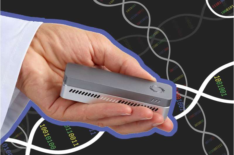
It cost nearly $3 billion to sequence the first human genome in 2001. Today, the cost to sequence a whole human genome is under $1000 and expected to reach about $100 soon. In addition, first- and second-generation sequencing systems were large, expensive, and designed for batch operation. Results would become available days or more after samples were taken. But new, lower-cost third-generation sequencing systems now exist, such as the Oxford Nanopore MinION, which can rapidly sequence individual samples and fit in the palm of your hand.

The human genome is made up of over three billion base pairs of DNA. To sequence a genome, the MinION employs small nanopores to divide a collected sample into billions of strands, called "reads."

"The MinION is a great handheld sequencing tool and is capable of rapidly sequencing biological data," says Reetuparna Das, an associate professor in CSE. "It takes the chemical sample, divides the DNA or RNA into strands, and sequences those strands into electrical signals, known as 'squiggles." However, it does not have the compute capability to analyze raw data in the field and quickly produce actionable results."

All that stands between us and real-time diagnosis is a computing system that can analyze the sequenced data and provide treatment and therapy recommendations before the patient even leaves the office.

The computing challenges

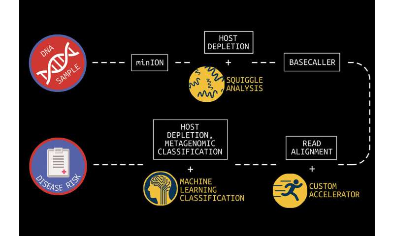
In what is known as secondary analysis, it is the job of a computing system to interpret squiggles as base pairs of DNA, a process which is known as basecalling. A base pair is essentially one rung on a DNA or RNA structure's ladder. Following that, the system must align the read data to genome reference data and then identify variants between the sample and the reference. The variant data of human genomes is used to identify a genetic disease marker. Sequencing is also used to identify pathogens by aligning DNA or RNA strands to a reference pathogen database and using metagenomic classification tools.
Precision health in the palm of your hand
U-M researchers are working to bring real-time diagnosis to healthcare providers through combined efforts in computer architecture and machine learning development. This graphic depicts the full pipeline necessary to get from DNA sample to actionable diagnosis. Each step is labeled in a white box, and the tools being developed at U-M to address that step below along with an illustration. The researchers use DNA data sequenced by the Oxford Nanopore MinION device. Credit: University of Michigan

And although this sounds straightforward, sequencing produces about GBs to TBs of data and the processing challenges are steep because of the precision, complexity, and scale of the task. Two multidisciplinary teams of researchers at U-M are working on approaches to overcome this hurdle.

Associate professor Reetuparna Das and professor Satish Narayanasamy, along with professor David Blaauw in Electrical and Computer Engineering, are leading a team funded by the National Science Foundation and the Kahn Foundation that is developing a hardware/software platform to accelerate next-generation genomic sequencing with a focus on pathogen detection and early cancer detection. In this effort, they are collaborating with associate professor of internal medicine and of microbiology and immunology Robert Dickson and assistant professor Carl Koschmann of pediatrics, as well as with associate professor Jenna Wiens, who is also a part of the second research team.

The second team, funded by the Kahn Foundation, is developing data acquisition and machine learning techniques to dramatically improve the prediction, treatment, and management of disease in aging populations. A key component of this effort is the use of machine learning to speed metagenomic analyses.

This large-scale interdisciplinary effort is a collaboration between researchers at Technion—Israel Institute of Technology, the Weizmann Institute, and U-M. The U-M researchers are led by Betsy Foxman, professor of epidemiology at the School of Public Health. Wiens, who is also a Co-Director of U-M Precision Health, is a Co-PI for the U-M research group.

An accelerated computing platform for genomic sequencing

Blaauw, Das, and Narayanasamy are focused on dramatically accelerating and optimizing the pipeline to process data from the MinION. The goal, say the researchers, is to reduce the time required to analyze a sequenced genome from hundreds of CPU hours to a matter of minutes.

"To realize the full potential of genomic sequencing," says Das, "computing power needs to increase by orders of magnitude."

The problem is, that's not possible under traditional processor roadmaps, where additional transistors and cores are packed ever more tightly into a processor for incremental processing gains. Added additional programming cores won't solve the problem either.

"Sustainable growth of processor performance is only possible with custom layers including hardware, software, and algorithms," says Das.

There are a number of areas of inefficiency that occur during secondary analysis which the team is addressing.

First, says Das, is the read alignment process, during which read data is aligned to genome reference data. Read alignment is composed of two steps: seeding and seed extension.
Precision health in the palm of your hand
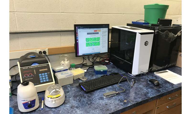
A faster pipeline for analyzing sequenced data: An Oxford Nanopore MinION in the researchers' lab enables rapid, mobile genome sequencing. U-M researchers are working to accelerate the efficiency of downstream genome and microbiome analysis. | Credit: Reetuparna Das

Seeding finds a set of candidate locations in the reference genome where a read can align. Possible matches are known as hits in the reference. In seed extension, for a read, the reference strings at the hit positions are matched at the read. With current technology, this takes hundreds of CPU hours for a whole genome.

For seeding, the researchers discovered a huge memory bandwidth bottleneck. They did hardware/software codesign and developed a new algorithm, data structure, and index that trades off memory capacity for memory bandwidth. They then built a custom accelerator that traverses the new index efficiently to find hits and seeds. The seeding algorithm has been released as open source software and is planned to be integrated with state of art alignment software from Broad Institute and Intel.

For seed extension, they built a systolic array that would in a few hundred cycles use approximate string matching to match read and reference data.

The researchers have developed a custom ASIC to eliminate the throughput bottleneck by using a pruning algorithm to optimize the alignment of DNA reads and candidate mutations and by reducing floating point computation by 43x when tested on real human data.

These enhancements and others have been mapped to custom hardware. This includes an accelerator for seed extension which achieves 2.46M reads/second, a ~1800x performance improvement, and a 27x smaller silicon footprint compared to a Xeon E5420 processor.

According to the researchers, when run on a high-end 56-core server in the Amazon cloud, their secondary analysis tools will take about six hours for whole genome sequencing. On an Amazon FPGA server, this reduces to about 20 minutes. When run on the researchers' custom hardware, processing time is about a minute.

The team has also developed techniques to optimize the read process for pathogen detection. One is to quickly analyze the beginning of a read to determine if it is host or pathogen material. If it is host, the remainder of the read can be skipped since it is only the pathogen material that is of interest. In addition, the researchers are often able to accomplish this host vs. pathogen differentiation using machine learning on squiggle data, without the need for resource-intensive basecalling.

Microbiome analysis to provide faster insights

When processing clinical samples, a fast data processing pipeline is key to the delivery of actionable insights.

"In clinical samples, much of the data—sometimes as much as 90%—can be host DNA, rather than microbial DNA," says Meera Krishnamoorthy, a Ph.D. student working with Wiens. "As a result, existing metagenomic classification tools store a lot of information about the host, and this can get computationally inefficient."

In collaboration with a team of researchers in Michigan Medicine and the School of Public Health, Wiens and Krishnamoorthy are working on in-silico machine learning approaches to host depletion, or the removal of host data reads, which will become a part of Das, Blaauw, and Narayanasamy's custom hardware. Their goal is to remove all of that host data before classification allowing downstream microbiome analyses to focus solely on microbial data. Existing host depletion methods are laboratory based and can be resource intensive to perform.

In contrast, Krishnamoorthy and Wiens' approach is computational and does not rely on large reference databases, but instead is based on a convolutional neural network. It takes as input read output by the basecaller and then after a series of convolutions and pooling steps outputs a prediction regarding whether or not the read pertains to the host. The proposed approach proposes to increase the efficiency of downstream analyses, enabling microbiome research that has the potential to transform future medical care.

Friday, January 08, 2021

 IPhone12 will stop your implantable defibrillator

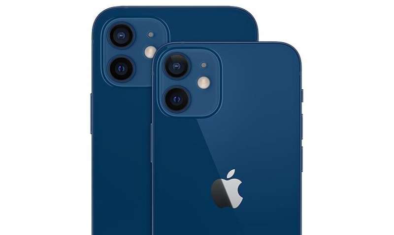
iPhone 12 and 12 Pro

In a recent paper in the journal Heart Rhythm, doctors describe how they turned off the potentially life-saving cardiac defibrillator function of an implanted Medtronic device simply by holding an iPhone 12 near it. The authors had nothing personal against Medtronic, or for that matter, against the new iPhone. The main reason they singled the phone out here was because it is compatible with some of the most advanced new technologies available for various magnetic-based communications and charging.

08 jan 2021--This technology, known as MagSafe, is basically harmless. It typically integrates charger, magnetometer and NFC reader into a compact package that depends on fairly decent alignment for efficient operation. The problem, at least for Medtronic, is the magnets that facilitate the positioning and docking. The iPhone 12, for example, has a ring of them around its central charging coil. In a nutshell, permanent magnets are never going away, they are simply a perfect solution to many gadget problems. Applications including securing cochlear implant links, joining cables and fastening wristbands now make extensive of use of strong, miniature magnets.

Unless companies like Medtronic get on board and move to smarter device configuration options, they will continue to butt heads with consumer devices—and they will continue to lose. Smarter options don't have to be expensive; just look at your cheap IR TV remote or ultrasonic receiver-emitter pair. These devices simply work. They use an uncomplicated code to make sure there is no interference from all the other ambient sources that are invariably present. A couple of secure ultrasonic bits superimposed on your basic 40 khz carrier waves is all that is really needed. It is likely that companies like Medtronic are working on solutions like this; for example, a Medtronic programming head of some sort can be had on Ebay at the moment for a mere $34.99.

In the larger scheme of things, having a handy iPhone, iWatch, fitbit, or even JUUL vape pen in your pocket to turn off inappropriate pulses or change stimulation modes is not such a bad thing, considering the alternative. Note that all these devices have accidentally toggled pacemakers. For example, if you have say, a standard issue Medtronic c Implantable Cardioverter Pacemaker or Resynchronization Defibrillator, they don't give you a tiny pen. Instead, you lug around their giant 3" diameter,5/8" thick donut magnet that gives a field of 90 Gauss at 1.5."

Perhaps now is a good time to look a little closer into what different implantable pacemaker/defibrillators actually do, and why inappropriately triggering them—or not triggering them, as the case may be—is undesirable. Inappropriate triggering is nothing unusual; it is, in fact, the central preoccupation for these devices. In other words, choosing when to force a contraction, or rhythm, and when to let the heart try to take some responsibility. There are different ways to implant, record from, and stimulate an ailing heart. You can do it externally to the heart chambers, inside the atrium, the ventricle, or both depending on the condition or pathology.

For example, with permanent chronic atrial fibrillation you might get by with a single atrial lead, while with intermittent or paroxysmal fibrillation, you likely want dual atrial and ventricular leads. In pacemaker vernacular, common control modes have names like AOO (asynchronous atrial pacing), VOO (asynchronous ventricular pacing), or DOO (asynchronous A+V pacing). More advanced modes, like DDD mode, have additional logical if-then control, something like the following: "dual-chamber anti bradycardia pacing; if atria fails to fire, it is paced. If the ventricle fails to fire after an atrial event (sensed or paced) the ventricle will be paced."

For the case of the accidental activation by a smartwatch, researchers replicated the misbehavior using a Medtronic Visia AF MRI S DF-1 single chamber ICD defibrillator. In the case of the accidental activation by a JUUL case, a man with a prolonged H-V interval had a dual-chamber Medtronic Evera MRI XT DR DDMB1D1. The reporting authors noted that although in this case, he was fine after reverting to magnet mode and halting emergency stimulation, there is clear potential for unintentional temporary programming and arrhythmic complications with these devices as they stand now.


More information: Joshua C. Greenberg, et al. Life Saving Therapy Inhibition by Phones Containing Magnets, Heart Rhythm. DOI: doi.org/10.1016/j.hrthm.2020.12.032

Wednesday, January 06, 2021

Memorable music could reduce anxiety amongst elderly during COVID vaccinations, says researcher

care home
Credit: Pixabay/CC0 Public Domain

Care providers should play music during the administration of coronavirus vaccinations to reduce feelings of anxiety in the elderly, researchers have said.

06 jan 2021--Research by Birmingham City University academic Mark Brill with Pendine Park Care Homes in Wales found that using music at the point of care can reduce stress, particularly for those living with dementia.

"Our research with care homes has shown how powerful music is in supporting people living with dementia, making care tasks easier, so it feels like a natural extension to use it during vaccination," explained Mark Brill, a senior lecturer in digital innovation at Birmingham City University.

Brill worked alongside Manchester Metropolitan University's Dr. Stuart Cunningham and Dr. Harry Whalley from the University for the Creative Arts in the wellbeing study.

Through published research, the team have shown that playing appropriate music during care activities can have a significant impact in managing anxiety and agitation and make care easier to provide.

The peer reviewed study showed that memorable songs can cut through anxiety, open up communication and create a relaxed atmosphere where effective care can take place.

"Care homes are under increased pressures, yet something as simple as music can bring relief all-round," added Mark. "We have prepared guides and support materials which would help make it easy for care staff and clinicians to use the technology during the vaccination process."


More information: Stuart Cunningham et al. Assessing Wellbeing in People Living with Dementia Using Reminiscence Music with a Mobile App (Memory Tracks): A Mixed Methods Cohort Study, Journal of Healthcare Engineering (2019). DOI: 10.1155/2019/8924273
Provided by Birmingham City University 

Saturday, January 02, 2021


Unique susceptibility to unique Sars-CoV-2 variants and vaccines

Unique susceptibility to unique Sars-CoV-2 variants and vaccines
Credit: Wikipedia

Individuals with different genetic variants in their immune system components often have very different immune responses to Sars-CoV-2. They also will have different responses to vaccines. By the same token, newly emerged variants in Sars-Cov-2 can elicit different immune responses in identical immune systems. In the larger reality we are now dealing, potential variation in all the above must be simultaneously considered.

02 Jan 2021--We recently discussed several sources of newly uncovered variation in immune genes that control susceptibility to Sars. For example, modern humans with throwback versions of the neanderthal gene DPP4, or the spike protein cleaving protease TMPRSS2 appear to be at high risk for severe Covid. Other variants, like a highly-expressed TMEM1B gene common in East Asians, or in genes of the heparin sulfate synthesis pathway, help explain the disproportionate severity of COVID-19 in some populations

Two papers, one recently published in Nature Immunology, and the other in Science, now extend the emerging gene list to include variations in the structure of the antibodies that are elicited by virus or vaccine. These variations include a particular kind of posttranslational modification of the anti-RBD (receptor binding domain) of the abundant IgG1 subclass known as afucosylation. What this essentially means is that for one reason or another, the afucosylated antibody is missing the addition of a fucose sugar molecule at a key structural location.

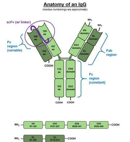
To better visualize the effect of this situation consider the above picture of the standard issue IgG antibody molecule. The variable portion, the arms of the Y shaped molecule, is known as the Fab region and it contains the epitopes that interact with the virus spike protein. The singular base region contains the constant Fc fragment, and incidentally, the glycosylated adornment hanging off of position N297 that potentially contains the fucose. In an almost fractal-like idiosyncrasy, this glycoform piece of kit also has a similar biantennary structure to the parent antibody, only on a much smaller scale, and with various sugars instead of amino acids. The fucose itself hangs off the stem portion of the aggregate glycoform, much as these glycans in turn hang on the antibody.

For anyone familiar with a related type of posttranslational modification known as ubiquitination, the N-glycan linkages form a kind of a code, much like the so-called ubiquitin code. Whereas the ubiquitin code consists of linear and branching subunits of variously phosphorylated or acetylated ubuitins, SUMOs, or NEDDs, the N-glycan code is composed of a heptasaccharide core which can be further extended with core fucose, terminal galactose (Gal), terminal sialic acid (Sia), and bisecting GlcNAc through selective enzymatic glycosylation reactions.

What the authors of these new papers found is that patients with severe forms of Sars-CoV-2 had increased likelihood of IgG1 antibodies with afucosylated Fc glycans, particularly in males. The net result of afucosylation proved to be enhanced interaction and binding with the immune activating Fcγ receptor, FcγRIIIA. This subsequently increases production of cytokines like interleukin-6 and TNF by monocytes. It should be noted that the canonical Fc receptors for human IgG include both activating (FcγRI, FcγRIIA, FcγRIIC, FcγRIIIA, and FcγRIIIB) and inhibitory (FcγRIIB) receptors. Most immune effector cells coexpress both activating and inhibiting FcγRs and therefore the net outcome of IgG interactions can often be predicted as the ratio of the binding affinities of a specific activating to inhibiting IgG receptors.

The way the authors figured this kind of stuff out was to first isolate the relevant IgG from a patient's serum using protein purification and tryptic digestion. Then nanoscale liquid chromatography coupled to tandem mass spectrometry is generally used to characterize the sites of potential glycosylation. But what the authors really wanted to get at was a more quantitative description of how much receptor binding is affected by afucosylation. To do this, various tried-and-true biophysical techniques including Surface Plasmon Resonance (SPR), Isothermal Titration Calorimetry (ITC), Microscale Thermophoresis (MST), and Biolayer Interferometry (BLI) are now available.

The authors of the Nature paper choose to use Biolayer Interferometry, which yielded graphs for binding (in nm units) as a function of time from which they could derive kinetic constants. BLI is an optical, label-free technique that analyzes the interference pattern of white light reflected from two surfaces. One is a layer of immobilized protein on the biosensor tip while the other is an internal reference. When a ligand immobilized on the biosensor tip surface binds to an analyte in solution there is a shift in the interference pattern that can be measured in real-time.

Afucosylated IgG was found to have a 20–40-fold increase in affinity to FcγRIIIa. This helps explain observed shifts in the front-line lung scavengers expressing FcγRIIIA, namely the alveolar macrophages, into an activated state of antibody-dependent cellular phagocytosis. While lowered Fc fucosylation is seen in the anti-Spike responses of the ARDS patients, other research has found that high levels of fucosylation are found in several kinds of cancer. In attempting to generate high-titer convalescent immunoglobulin treatments, it is likely important that plasma enriched in fucosylated anti-SARS-CoV-2 antibodies should be used. Although afucosylated IgG formed against viruses generally mediate stronger FcγRIIIa responses, they will often amplify cytokine storm and immune pathology.

While variants in the many genes that help build glycan chains, like for example the fucosyltransferase FUT2, might be expected to play a role in patient responses, perhaps an ever more pressing concern is differences in the antibody response to vaccines. In a brief spate of good news, the actual sequence of the new Pfizer mRNA vaccine was generously released unto the masses. The quickest to respond and attempt to decode this revelation was a guy named Bert Hubert, who quickly broke it down for us.

The main difference in the vaccine code is that uracil has been replaced by 1-methyl-3'-pseudouridylyl, which is labelled as Ψ. Although Ψ does not arouse the ire of our immune systems, it is still accepted as a normal uracil by the translation, transcription, and replication machinery of the cell. The other curious features are the substitutions of two prolines to stabilize the spike structure, and the deployment of special 5' and 3' untranslated regions before and after the main spike sequence. Bert has promised to release a second deep dive into the sequence in a few days, which is no doubt anxiously awaited by many.


More information: Saborni Chakraborty et al. Proinflammatory IgG Fc structures in patients with severe COVID-19, Nature Immunology (2020). DOI: 10.1038/s41590-020-00828-7

Mads Delbo Larsen et al. Afucosylated IgG characterizes enveloped viral responses and correlates with COVID-19 severity, Science (2020). DOI: 10.1126/science.abc8378


A year of blursdays: how coronavirus distorted our sense of time in 2020

A year of blursdays: how coronavirus distorted our sense of time in 2020
Credit: StunningArt/Shutterstock

Does it feel like 2020 went on forever? Did lockdown drag, and can you even remember how you spent your time when you weren't living under coronavirus restrictions? You are not alone. For many, 2020 has been the year in which the constancy of time was lost to the upheaval of coronavirus.

02 jan 2021--Objectively, time passes at a constant, linear rate. Subjectively, however, time waxes and wanes with our activities and emotions. Sometimes, it flies by, other times it drags so slowly that it almost stands still.

This is backed up by research I conducted in April, which explored how the early months of the coronavirus pandemic had affected people's experiences of the passage of time. Of particular interest was how quickly time felt like it was passing during lockdown in comparison to "normal" (that long-ago time before lockdown).

I surveyed 604 people about how quickly time felt it was passing that day and that week in comparison to before the lockdown. Participants also answered questions about their mood, family life and how busy they were to give context on the factors, which made time more likely to speed up or slow down for different people.

Tempus fugit?

My results showed that there was widespread distortion time during lockdown, with more than 80% of people reporting that time felt like it was passing differently. But lockdown did not distort time in the same way for everyone. Instead, time sped up during lockdown for 40% of people and slowed down for the remaining 40%.

Why was this? My analysis suggests that the perceived speed of time during the day was affected by a person's age, how satisfied they were with their level of social interaction, how stressed they were and how busy they were. In general, the days passed more quickly for younger people who were socially satisfied, busy and experiencing low levels of stress. Conversely, the day passed more slowly for older people, particularly those over the age of 60, who were socially dissatisfied, stressed and lacking tasks to occupy them.

Similar patterns were observed for the subjective speed of the week. A fast week was associated with being younger and more socially satisfied, whereas a slow week was associated with being older and less socially satisfied.

A second unpublished study I conducted during the November lockdown revealed that, of the 851 people surveyed, more than 75% experienced distortion to time and 55% reported that the start of the first lockdown felt longer than eight months ago. A slower second lockdown was associated with shielding, dissatisfaction with social interaction and greater depression and boredom.

The UK is not alone in its loss of time during lockdown. Studies conducted in FranceItaly and Argentina also show widespread distortion to the passage of time during periods of strict COVID-19 restrictions.

Unlike in the UK, in France and Italy lockdown passed more slowly than normal for most people rather than being split 40/40 as in my April study. As in the UK, however, boredom was an important predictor of time slowing down in Italy and in France. In France, time also passed more slowly with increasing sadness.

Emotions and time

Why does being older, bored, stressed and socially dissatisfied make time pass more slowly? This question is difficult to answer.

Unlike other senses, we don't have an obvious organ for time. Instead, time is experienced as part of other sensory inputs, such as sight and hearing, and this has made it difficult to identify precisely how the brain processes it.

One possibility is that when we are bored and socially dissatisfied we have lots of spare cognitive capacity and that we then use some of that capacity to increase our monitoring of time. This increased monitoring then results in time passing more slowly than normal, simply because we are more aware of time than normal. Another possibility is that the emotional consequence of lockdown altered the way the brain processes time.

In particular, the negative emotions associated with isolation, boredom, sadness and stress may have contributed to a slowing of time. However, inconsistent effects of depression and anxiety across studies suggests that the effect of emotion on time is complex.

So what of 2021? Will time regain its regular rhythm? It is difficult to say. With the first vaccines currently being deployed, we maybe more hopeful than ever that normality is just around the corner. The reality may be that normality is many months away.

Regardless, while we can't change the actual time it takes for the vaccination programme to be completed, there are some things which we can do to speed up the wait. By keeping busy, minimising stress, engaging in as much face-to-face or online social interaction as we can and by reducing our stress levels, we can help the journey back to normality pass more quickly than normal.


Provided by The Conversation 

Thursday, December 17, 2020

 LED lights found to kill coronavirus

led
Credit: CC0 Public Domain

Researchers from Tel Aviv University (TAU) have proven that the coronavirus can be killed efficiently, quickly, and cheaply using ultraviolet (UV) light-emitting diodes (UV-LEDs). They believe that the UV-LED technology will soon be available for private and commercial use.

17 december 2020--This is the first study conducted on the disinfection efficiency of UV-LED irradiation at different wavelengths or frequencies on a virus from the family of coronaviruses. The study was led by Professor Hadas Mamane, Head of the Environmental Engineering Program at TAU's School of Mechnical Engineering, Iby and Aladar Fleischman Faculty of Engineering. The article was published in November 2020 issue of the Journal of Photochemistry and Photobiology B: Biology.

"The entire world is currently looking for effective solutions to disinfect the coronavirus," said Professor Mamane. "The problem is that in order to disinfect a bus, train, sports hall, or plane by chemical spraying, you need physical manpower, and in order for the spraying to be effective, you have to give the chemical time to act on the surface. Disinfection systems based on LED bulbs, however, can be installed in the ventilation system and air conditioner, for example, and sterilize the air sucked in and then emitted into the room.

"We discovered that it is quite simple to kill the coronavirus using LED bulbs that radiate ultraviolet light," she explained. "We killed the viruses using cheaper and more readily available LED bulbs, which consume little energy and do not contain mercury like regular bulbs. Our research has commercial and societal implications, given the possibility of using such LED bulbs in all areas of our lives, safely and quickly."

The researchers tested the optimal wavelength for killing the coronavirus and found that a length of 285 nanometers (nm) was almost as efficient in disinfecting the virus as a wavelength of 265 nm, requiring less than half a minute to destroy more than 99.9% of the coronaviruses. This result is significant because the cost of 285 nm LED bulbs is much lower than that of 265 nm bulbs, and the former are also more readily available.

Eventually, as the science develops, the industry will be able to make the necessary adjustments and install the bulbs in robotic systems or air conditioning, vacuum, and water systems, and thereby be able to efficiently disinfect large surfaces and spaces. Professor Mamane believes that the technology will be available for use in the near future.

It is important to note that it is very dangerous to try to use this method to disinfect surfaces inside homes. To be fully effective, a system must be designed so that a person is not directly exposed to the light.

In the future, the researchers will test their unique combination of integrated damage mechanisms and more ideas they recently developed on combined efficient direct and indirect damage to bacteria and viruses on different surfaces, air, and water.


More information: Yoram Gerchman et al, UV-LED disinfection of Coronavirus: Wavelength effect, Journal of Photochemistry and Photobiology B: Biology (2020). DOI: 10.1016/j.jphotobiol.2020.112044
Provided by Tel Aviv University 

Smartphone camera used to diagnose viral infections

Smartphone camera used to diagnose various viral infections
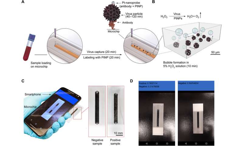
Three-dimensional schematic of the CNN–nanoparticle-enabled smartphone for virus detection. The detection process comprises three main steps. (A) Virus capture and labeling using Pt-nanoprobes. The samples are loaded into microchips modified with mAbs against the virus envelope protein and incubated to allow the capture of target virus for 20 min. The captured virus particles are then labeled with Pt-nanoprobes within 20 min. Each step of on-chip virus capture and labeling is followed by a washing step using 10 mM phosphate buffer (pH 7.4) (B) Catalyzer solution containing hydrogen peroxide (H2O2) is added to the chip and incubated for 10 min. In the presence of the captured virus, Pt-nanoprobe complex bubbles (oxygen) are formed because of the catalytic activity of PtNPs in contact with H2O2. Scale bar is set on the basis of the microchip dimensions. (C) The on-chip bubble signal is detected using the CNN–nanoparticle-enabled smartphone (NES). Photo credit: Mohamed S. Draz, Brigham and Women’s Hospital. (D) Screenshots of the results shown by the CNN-NES. Photo credit: Mohamed S. Draz, Brigham and Women’s Hospital. Credit: Science Advances (2020). DOI: 10.1126/sciadv.abd5354

A team of researchers at Brigham and Women's Hospital, Harvard Medical School, has developed a way to use a smartphone camera to test for viral infections. In their paper published in the journal Science Advances, the group describes their system, which also involves the use of an external catalytic microchip device and a smartphone system that uses a trained deep learning algorithm.

17 december 2020--As the pandemic has gripped the world for most of this year, scientists have been looking for ways to slow the spread of the next one. In this new effort, the team in Massachusetts has developed a smartphone-based system that can be used by non-medical people to test for a variety of viral infections.

The system is made up of a smartphone, an external catalytic microchip device and software. Body fluid samples are placed into a channel on the catalytic microchip device, which is then doused with a small amount of hydrogen peroxide. The resulting reaction leads to the formation of bubbles. The bubbles develop in unique patterns based in part on viruses in the fluid sample. The user points their smartphone camera at the bubbling sample and launches the deep-learning algorithm that has already been trained to identify the patterns and thereby recognize the presence of viruses. The whole process takes approximately 50 minutes. The researchers have thus far taught their system to recognize just three viruses, Zika and hepatitis B and C. But testing shows the system to be 99% accurate. They note that their system is more portable and cost effective than other solutions in the works.

The researchers suggest that their system could be rapidly trained to recognize new viruses if the need arises, and the catalytic microchip device could be sent to hot spots in the future. Such technology, the researchers suggest, could help to stop future pandemics if used widely. The researchers also note that the system could be immediately useful in infection prone areas lacking testing labs, such as third world countries.


More information: Mohamed S. Draz et al. Virus detection using nanoparticles and deep neural network–enabled smartphone system, Science Advances (2020). DOI: 10.1126/sciadv.abd5354


Sunday, December 13, 2020

Existing antihistamine drugs show effectiveness against COVID-19 virus in cell testing

Existing antihistamine drugs show effectiveness against COVID-19 virus in cell testing
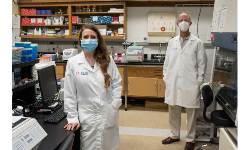
Leah Reznikov, Ph.D., and David Ostrov, Ph.D., are shown in Reznikov’s lab. Credit: UF Health/Jesse Jones

Three common antihistamine medications have been found in preliminary tests to inhibit infection of cells by the coronavirus that causes COVID-19, University of Florida Health researchers have found.

13 dec 2020--Their findings, based on laboratory tests of cells and a detailed analysis of nearly a quarter-million California patients' medical records, are published today in the journal Biochemical and Biophysical Research Communications. The data may support the launch of a randomized, controlled clinical trial to determine whether the specific antihistamines can treat or even prevent COVID-19 in humans, the researchers said.

Earlier this year, Leah Reznikov, Ph.D., an assistant professor of physiological sciences in the UF College of Veterinary Medicine and the study's principal investigator on the study, began collaborating with David A. Ostrov, Ph.D., an immunologist and associate professor in the UF College of Medicine's department of pathology, immunology and laboratory medicine. They and other colleagues set out to identify approved drugs that can interfere with the way the SARS-CoV-2 virus binds to cells. Blocking that connection essentially locks a cellular doorway that inhibits the virus's transmission to the respiratory system.

"We discovered epidemiological data showing that the usage of specific drugs was associated with a reduced likelihood of testing positive for SARS-CoV-2, the virus that causes COVID-19. We then found that these specific drugs exhibited direct antiviral activity against SARS-CoV-2 in the lab," Ostrov said.

While there is an association between the medications and infection rates, the researchers stressed there is much more to be learned and no cause and effect has been formally established.

"The fact that these drugs actually inhibit the virus in the lab does not necessarily mean that they will inhibit it actively in people—but they might," Ostrov said.

To establish their findings, the collaborators focused on angiotensin-converting enzyme-2, or ACE2, a "gateway" protein the virus uses to invade human cells. Working with a colleague at the University of California San Francisco, they analyzed medical records of nearly a quarter million California patients. People age 61 and older who had used certain antihistamines were less likely to test positive for the SARS-CoV-2 virus than those who did not take the medications, the researchers found.

Next, the researchers tested this group of antihistamines for their ability to inhibit the coronavirus in a combination of human and primate cells. Three of the drugs—hydroxyzine, diphenhydramine and azelastine—showed direct, statistically significant antiviral effects on the SARS-CoV-2 virus.

Hydroxyzine, sold as Atarax, and the nasal spray azelastine are prescription medications while diphenhydramine is sold over-the-counter as Benadryl, a treatment for cold and allergy symptoms.

The drugs were tested at different concentrations to measure how much is required to inhibit the virus.

While the findings are encouraging, Ostrov cautions against self-medicating with antihistamines as a COVID-19 prevention or treatment. So-called "off-label" use of medications should only take place after a detailed consultation with a physician, he said.

Among the three medications, azelastine was found to inhibit the SARS-CoV-2 virus at a dose that was smaller than the amount prescribed as a nasal spray. The other two antihistamines required higher drug concentrations than currently recommended dosing levels to achieve antiviral activity in cells. That doesn't make diphenhydramine any less of a potential COVID-19 therapy for now, especially considering its ubiquity and over-the-counter status, Ostrov said. Clinical trials will be necessary to establish the drugs' effectiveness in prevention, early treatment and as a secondary therapy for severe COVID-19.

Reznikov said the data suggest these three antihistamines may work by either disrupting the virus's interactions with ACE2 or by binding with another protein that may interfere with viral replication. The protein, known as a sigma receptor, is part of a cell's communications network.

Reznikov approached Ostrov in March with a hypothesis and an idea: Drugs that bind ACE2 could change disease outcomes, so she asked Ostrov to develop a list of small-molecule drug candidates.

She then pursued a strategy to screen the drugs against virus isolates of SARS-CoV-2 with Michael Norris, Ph.D., an assistant research professor of medical geography in the department of geography and the UF Emerging Pathogens Institute. In May, Reznikov was included in the UF Clinical and Translational Science Institute's Rapid-Response Translational Research Funding initiative to facilitate this project.

Although the findings are intriguing, Reznikov and Ostrov note there is still much to be learned about the mechanisms of how antihistamines interact with the SARS-CoV-2 virus.

Because of the urgency of the COVID-19 pandemic, Ostrov said there is a possibility that the antihistamine candidates could go directly to human clinical trials without first being tested in animal models. That is the case with famotidine, an antihistamine and antacid that is undergoing clinical trials elsewhere as a therapy for severely ill COVID-19 patients.


More information: Leah R. Reznikov et al. Identification of antiviral antihistamines for COVID-19 repurposing, Biochemical and Biophysical Research Communications (2020). DOI: 10.1016/j.bbrc.2020.11.095
Provided by University of Florida 

Friday, December 11, 2020

Gut microbiota plays a role in brain function and mood regulation

Gut microbiota plays a role in brain function and mood regulation
Credit: Pascal Marseaud

Depression is a mental disorder that affects more than 264 million people of all ages worldwide. Understanding its mechanisms is vital for the development of effective therapeutic strategies. Scientists from the Institut Pasteur, Inserm and the CNRS recently conducted a study showing that an imbalance in the gut bacterial community can cause a reduction in some metabolites, resulting in depressive-like behaviors. These findings, which show that a healthy gut microbiota contributes to normal brain function, were published in Nature Communications on December 11, 2020.

11 december 2020--The bacterial population in the gut, known as the gut microbiota, is the largest reservoir of bacteria in the body. Research has increasingly shown that the host and the gut microbiota are an excellent example of systems with mutually beneficial interactions. Recent observations also revealed a link between mood disorders and damage to the gut microbiota. This was demonstrated by a consortium of scientists from the Institut Pasteur, the CNRS and Inserm, who identified a correlation between the gut microbiota and the efficacy of fluoxetine, a molecule frequently used as an antidepressant. But some of the mechanisms governing depression, the leading cause of disability worldwide, remained unknown.

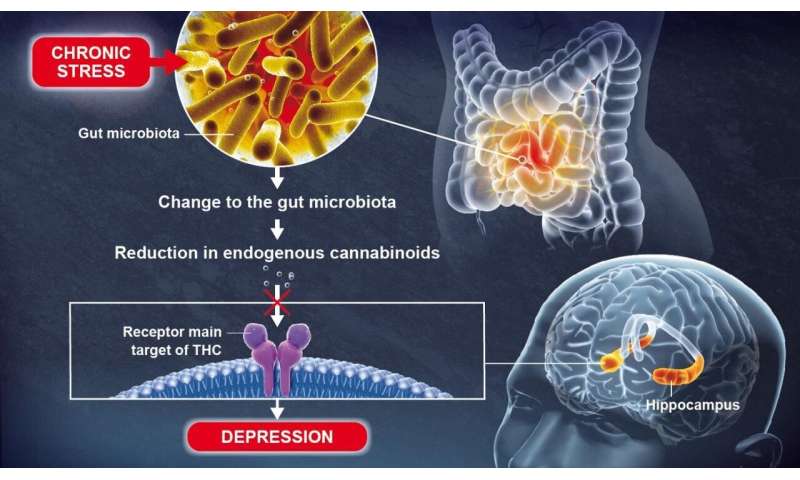
Using animal models, scientists recently discovered that a change to the gut microbiota brought about by chronic stress can lead to depressive-like behaviors, in particular by causing a reduction in lipid metabolites (small molecules resulting from metabolism) in the blood and the brain.

These lipid metabolites, known as endogenous cannabinoids (or endocannabinoids), coordinate a communication system in the body which is significantly hindered by the reduction in metabolites. Gut microbiota plays a role in brain function and mood regulation

Endocannabinoids bind to receptors that are also the main target of THC, the most widely known active component of cannabis. The scientists discovered that an absence of endocannabinoids in the hippocampus, a key brain region involved in the formation of memories and emotions, resulted in depressive-like behaviors.

The scientists obtained these results by studying the microbiotas of healthy animals and animals with mood disorders. As Pierre-Marie Lledo, Head of the Perception and Memory Unit at the Institut Pasteur (CNRS/Institut Pasteur) and joint last author of the study, explains: "Surprisingly, simply transferring the microbiota from an animal with mood disorders to an animal in good health was enough to bring about biochemical changes and confer depressive-like behaviors in the latter."

The scientists identified some bacterial species that are significantly reduced in animals with mood disorders. They then demonstrated that an oral treatment with the same bacteria restored normal levels of lipid derivatives, thereby alleviating the depressive-like behaviors. These bacteria could therefore serve as an antidepressant. Such treatments are known as "psychobiotics".

"This discovery shows the role played by the gut microbiota in normal brain function," continues Gérard Eberl, Head of the Microenvironment and Immunity Unit (Institut Pasteur/Inserm) and joint last author of the study. If there is an imbalance in the gut bacterial community, some lipids that are vital for brain function disappear, encouraging the emergence of depressive-like behaviors. In this particular case, the use of specific bacteria could be a promising method for restoring a healthy microbiota and treating mood disorders more effectively.


More information: Grégoire Chevalier et al, Effect of gut microbiota on depressive-like behaviors in mice is mediated by the endocannabinoid system, Nature Communications (2020). DOI: 10.1038/s41467-020-19931-2
Provided by Pasteur Institute 

Saturday, December 05, 2020

 How are older adults coping with the mental health effects of COVID-19?

How are older adults coping with the mental health effects of COVID-19?
McLean researchers indicate that older adults may be withstanding the mental health strains of the COVID-19 pandemic better than other age groups. Credit: McLean Hospital

Older adults are especially vulnerable to the effects of the COVID-19 pandemic—with higher risks of severe complications and death, and potentially greater difficulties accessing care and adapting to technologies such as telemedicine. A viewpoint article published in the Journal of the American Medical Association notes that there's also a concern that isolation during the pandemic could be more difficult for older individuals, which could exacerbate existing mental health conditions. Information gathered over the past several months suggests a much more nuanced picture, however.

05 december 2020--"Over the spring and summer of 2020, we were struck by a number of individual studies from all over the world that reported a consistent theme: Older adults, as a group, appeared to be withstanding the strains on mental health from the pandemic better than all other age groups," said lead author Ipsit Vahia, MD, medical director of the Geriatric Psychiatry Outpatient Services and the Institute for Technology in Psychiatryat McLean Hospital. "In this article, we highlight some of these studies and discuss resilience in older adults and what factors may be driving it."

Resilience may reflect an interaction among internal factors—such as an individual's stress response, cognitive capacity, personality traits, and physical health—and external resources like social connections and financial stability. For older adults experiencing isolation during the pandemic, having more meaningful relationships seems to be more important than having more interactions with others, and maintaining these relationships may require the use of technology to connect with loved ones.

Resilience can be supported through increased physical activity, enhanced compassion and emotional regulation, and greater social connectivity. Technology can play an important role in achieving these. "It can help maintain social connectivity, provide access to care via telemedicine, and also facilitate a range of other activities that may help cope with isolation," said Vahia. "It is increasingly becoming important for clinicians to assess patients' access and proficiency with technology as a part of care."

The authors stressed that although findings from the early months of the pandemic are encouraging and provide cause for cautious optimism, they may not reflect individual realities. "Older adults are a highly diverse group, and each person's response to the stresses of the pandemic depends on a unique set of circumstances," Vahia explained. "In addition, the current studies may not reflect specific high-risk populations with unique stressors, such as those living in underserved areas or those suffering with dementia or caregivers for people with dementia."

Importantly, the pandemic continues without a defined timeline or clear end in sight. The longer-term effects of COVID-19 on older adults' mental health, especially in countries with very high rates of disease, are unclear.


More information: Ipsit V. Vahia et al, Older Adults and the Mental Health Effects of COVID-19, JAMA (2020). DOI: 10.1001/jama.2020.21753
Provided by McLean Hospital 

Tuesday, December 01, 2020

Older adults with dementia exhibit financial 'symptoms' up to six years before diagnosis

dementia
Credit: CC0 Public Domain

A new study led by researchers at the Johns Hopkins Bloomberg School of Public Health and the Federal Reserve Board of Governors found that Medicare beneficiaries who go on to be diagnosed with dementia are more likely to miss payments on bills as early as six years before a clinical diagnosis.

01 december 2020--The study also found that beneficiaries diagnosed with dementia who had a lower educational status missed payments on bills beginning as early as seven years before a clinical diagnosis as compared to 2.5 years prior to a diagnosis for beneficiaries with higher educational status.

The study, which included researchers from the University of Michigan Medical School, also found that these missed payments and other adverse financial outcomes lead to increased risk of developing subprime credit scores starting 2.5 years before a dementia diagnosis. Subprime credit scores fall in the fair and lower range.

The findings, published online November 30 in JAMA Internal Medicine, suggest that financial symptoms such as missing payments on routine bills could be used as early predictors of dementia and highlight the benefits of earlier detection.

"Currently there are no effective treatments to delay or reverse symptoms of dementia," says lead author Lauren Hersch Nicholas, Ph.D., associate professor in the Department of Health Policy and Management at the Bloomberg School. "However, earlier screening and detection, combined with information about the risk of irreversible financial events, like foreclosure and repossession, are important to protect the financial well-being of the patient and their families."

The analysis found that the elevated risk of payment delinquency with dementia accounted for 5.2 percent of delinquencies among those six years prior to diagnosis, reaching a maximum of 17.9 percent nine months after diagnosis. Rates of elevated payment delinquency and subprime credit risk persisted for up to 3.5 years after beneficiaries received dementia diagnoses, suggesting an ongoing need for assistance managing money.

Dementia, identified as diagnostic codes for Alzheimer's Disease and related dementias in the study, is a progressive brain disorder that slowly diminishes memory and cognitive skills and limits the ability to carry out basic daily activities, including managing personal finances. About 14.7 percent of American adults over the age of 70 are diagnosed with the disease. The onset of dementia can lead to costly financial errors, irregular bill payments, and increased susceptibility to financial fraud.

For their study, the researchers linked de-identified Medicare claims and credit report data. They analyzed information on 81,364 Medicare beneficiaries living in single-person households, with 54,062 never receiving a dementia diagnosis between 1999 and 2014 and 27,302 with a dementia diagnosis during the same period. The researchers compared financial outcomes spanning 1999 to 2018 of those with and without a clinical diagnosis of dementia for up to seven years prior to a diagnosis and four years following a diagnosis. The researchers focused on missing payments for one or more credit accounts that were at least 30 days past due, and subprime credit scores, indicative of an individual's risk of defaulting on loans based on credit history.

To determine whether the financial symptoms observed were unique to dementia, the researchers also compared financial outcomes of missed payments and subprime credit scores to other health outcomes including arthritis, glaucoma, heart attacks, and hip fractures. They found no association of increased missed payments or subprime credit scores prior to a diagnosis for arthritis, glaucoma, or a hip fracture. No long-term associations were found with heart attacks.

"We don't see the same pattern with other health conditions," says Nicholas. "Dementia was the only medical condition where we saw consistent financial symptoms, especially the long period of deteriorating outcomes before clinical recognition. Our study is the first to provide large-scale quantitative evidence of the medical adage that the first place to look for dementia is in the checkbook."


More information: Lauren Hersch Nicholas et al. Financial Presentation of Alzheimer Disease and Related Dementias, JAMA Internal Medicine (2020). DOI: 10.1001/jamainternmed.2020.6432
Journal information: JAMA Internal Medicine