Saturday, October 30, 2021


New study provides robust evidence that COVID-19 is a seasonal infection

covid mask leaves
Credit: Unsplash/CC0 Public Domain

A new study led by the Barcelona Institute for Global Health (ISGlobal) provides robust evidence that COVID-19 is a seasonal infection linked to low temperatures and humidity, much like seasonal influenza. The results, published in Nature Computational Science, also support the considerable contribution of airborne SARS-CoV-2 transmission and the need to shift to measures that promote "air hygiene."

A key question regarding SARS-CoV-2 is whether it is behaving, or will behave, as a seasonal virus like influenza, or whether it will be equally transmitted during any time of the year. A first theoretical modeling study suggested that climate was not a driver in COVID-19 transmission, given the high number of susceptible individuals with no immunity to the virus. However, some observations suggested that the initial propagation of COVID-19 in China occurred in a latitude between 30 and 50 degrees N, with low humidity levels and low temperatures (between 5 degrees and 11 degrees C).

"The question of whether COVID-19 is a genuine seasonal disease becomes increasingly central, with implications for determining effective intervention measures," explains Xavier Rodó, director of the Climate and Health program at ISGlobal and coordinator of the study. To answer this question, Rodó and his team first analyzed the association of temperature and humidity in the initial phase of SARS-CoV-2 spread in 162 countries across five continents, before changes in human behavior and public health policies were put into place. The results show a negative relationship between the transmission rate (R0) and both temperature and humidity at the global scale: higher transmission rates were associated with lower temperatures and humidity.

The team then analyzed how this association between climate and disease evolved over time, and whether it was consistent at different geographical scales. For this, they used a statistical method that was specifically developed to identify similar patterns of variation (i.e. a pattern-recognition tool) at different windows of time. Again, they found a strong negative association for short time windows between disease (number of cases) and climate (temperature and humidity), with consistent patterns during the first, second and third waves of the pandemic at different spatial scales: worldwide, countries, down to individual regions within highly affected countries (Lombardy, Thüringen and Catalonia) and even to the city level (Barcelona).

The first epidemic waves waned as temperature and humidity rose, and the second wave rose as temperatures and humidity fell. However, this pattern was broken during summertime in all continents. "This could be explained by several factors, including mass gatherings of young people, tourism, and air conditioning, among others," explains Alejandro Fontal, researcher at ISGlobal and first author of the study.

When adapting the model to analyze transient correlations at all scales in countries in the Southern Hemisphere, where the virus arrived later, the same negative correlation was observed. The climate effects were most evident at temperatures between 12 degrees and 18 degrees C and humidity levels between 4 and 12 g/m3, although the authors warn that these ranges are still indicative, given the short records available.

Finally, using an epidemiological model, the research team showed that incorporating temperature into the transmission rate works better for predicting the rise and fall of the different waves, particularly the first and third ones in Europe. "Altogether, our findings support the view of COVID-19 as a true seasonal low-temperature infection, similar to influenza and to the more benign circulating coronaviruses," says Rodó.

This seasonality could contribute importantly to the transmission of SARS-CoV-2, since low humidity conditions have been shown to reduce the size of aerosols, and thereby increase airborne transmission of seasonal viruses such as influenza. "This link warrants an emphasis on 'air hygiene' through improved indoor ventilation as aerosols are capable to persist suspended for longer times," says Rodó, and highlights the need to include meteorological parameters in the evaluation and planning of control measures.


More information: Xavier Rodó, Climatic signatures in the different COVID-19 pandemic waves across both hemispheres, Nature Computational Science (2021). DOI: 10.1038/s43588-021-00136-6. www.nature.com/articles/s43588-021-00136-6
Provided by Barcelona Institute for Global Health 


How many people get 'long COVID?' More than half, researchers find

covid
Credit: Pixabay/CC0 Public Domain

More than half of the 236 million people who have been diagnosed with COVID-19 worldwide since December 2019 will experience post-COVID symptoms—more commonly known as "long COVID"—up to six months after recovering, according to Penn State College of Medicine researchers. The research team said that governments, health care organizations and public health professionals should prepare for the large number of COVID-19 survivors who will need care for a variety of psychological and physical symptoms.

30 oct 2021--During their illnesses, many patients with COVID-19 experience symptoms, such as tiredness, difficulty breathing, chest pain, sore joints and loss of taste or smell.

Until recently, few studies have evaluated patients' health after recovering from the coronavirus. To better understand the short- and long-term health effects of the virus, the researchers examined worldwide studies involving unvaccinated patients who recovered from COVID-19. According to the findings, adults, as well as children, can experience several adverse health issues for six months or longer after recovering from COVID-19.

The researchers conducted a systematic review of 57 reports that included data from 250,351 unvaccinated adults and children who were diagnosed with COVID-19 from December 2019 through March 2021. Among those studied, 79% were hospitalized, and most patients (79%) lived in high-income countries. Patients' median age was 54, and the majority of individuals (56%) were male.

The researchers analyzed patients' health post-COVID during three intervals at one month (short-term), two to five months (intermediate-term) and six or more months (long-term).

According to the findings, survivors experienced an array of residual health issues associated with COVID-19. Generally, these complications affected a patient's general well-being, their mobility or organ systems. Overall, one in two survivors experienced long-term COVID manifestations. The rates remained largely constant from one month through six or more months after their initial illness. 

The investigators noted several trends among survivors, such as:

  • General well-being: More than half of all patients reported weight loss, fatigue, fever or pain.
  • Mobility: Roughly one in five survivors experienced a decrease in mobility.
  • Neurologic concerns: Nearly one in four survivors experienced difficulty concentrating.
  • Mental health disorders: Nearly one in three patients were diagnosed with generalized anxiety disorders.
  • Lung abnormalities: Six in ten survivors had chest imaging abnormality and more than a quarter of patients had difficulty breathing.
  • Cardiovascular issues: Chest pain and palpitations were among the commonly reported conditions.
  • Skin conditions: Nearly one in five patients experienced hair loss or rashes.
  • Digestive issues: Stomach pain, lack of appetite, diarrhea and vomiting were among the commonly reported conditions.

"These findings confirm what many health care workers and COVID-19 survivors have been claiming, namely, that adverse health effects from COVID-19 can linger," said co-lead investigator Vernon Chinchilli, chair of the Department of Public Health Sciences. "Although previous studies have examined the prevalence of long COVID symptoms among patients, this study examined a larger population, including people in high-, middle- and low-income countries, and examined many more symptoms. Therefore, we believe our findings are quite robust given the available data."

"The burden of poor health in COVID-19 survivors is overwhelming," said co-lead investigator Dr. Paddy Ssentongo, assistant professor at the Penn State Center for Neural Engineering. "Among these are the mental health disorders. One's battle with COVID doesn't end with recovery from the acute infection. Vaccination is our best ally to prevent getting sick from COVID-19 and to reduce the chance of long-COVID even in the presence of a breakthrough infection."

The mechanisms by which COVID-19 causes lingering symptoms in survivors are not fully understood. These symptoms could result from immune-system overdrive triggered by the virus, lingering infection, reinfection or an increased production of autoantibodies (antibodies directed at their own tissues). The SARS-CoV-2 virus, the agent that causes COVID-19, can access, enter and live in the nervous system. As a result, nervous system symptoms such as taste or smell disorders, memory impairment and decreased attention and concentration commonly occur in survivors. 

"Our study was not designed to confirm COVID-19 as the sole cause of these symptoms. It is plausible that symptoms reported by patients in some of the studies examined were due to some other causes," said Ssentongo.

According to the researchers, early intervention will be critical for improving the quality of life for many COVID-19 survivors. They said that in the years ahead, health care providers will likely see an influx of patients with psychiatric and cognitive problems, such as depression, anxiety or post-traumatic stress disorder, who were otherwise healthy before their COVID-19 infection. Based on these findings, health care providers should plan and allocate resources accordingly in order to effectively monitor and treat these conditions.

The research team noted that these long-term health conditions may cause increased demand for medical care and could overwhelm health care systems, particularly in low- and middle-income countries. They said the findings from this study could help shape treatment plans to improve care for COVID-19 patients and establish integrated evidence-based clinical management for those affected.

"Since survivors may not have the energy or resources to go back and forth to their health care providers, one-stop clinics will be critical to effectively and efficiently manage patients with long COVID," Ssentongo said. "Such clinics could reduce medical costs and optimize access to care, especially in populations with historically larger health care disparities."

Th research was published in JAMA Network Open.

More information: Destin Groff et al, Short-term and Long-term Rates of Postacute Sequelae of SARS-CoV-2 Infection: A Systematic Review,
JAMA Netw Open (2021). DOI: 10.1001/jamanetworkopen.2021.28568
Provided by Pennsylvania State University 


Alzheimer's and COVID-19 share a genetic risk factor

gene
Credit: Pixabay/CC0 Public Domain

An anti-viral gene that impacts the risk of both Alzheimer's disease and severe COVID-19 has been identified by a UCL-led research team.

30 oct 2021--The researchers estimate that one genetic variant of the OAS1 gene increases the risk of Alzheimer's disease by about 3-6% in the population as a whole, while related variants on the same gene increase the likelihood of severe COVID-19 outcomes.

The findings, published in Brain, could open the door for new targets for drug development or tracking disease progression in either disease, and suggest that treatments developed could be used for both conditions. The findings also have potential benefits for other related infectious conditions and dementias.

Lead author Dr. Dervis Salih (UCL Queen Square Institute of Neurology and UK Dementia Research Institute at UCL) said: "While Alzheimer's is primarily characterized by harmful build-up of amyloid protein and tangles in the brain, there is also extensive inflammation in the brain that highlights the importance of the immune system in Alzheimer's. We have found that some of the same immune system changes can occur in both Alzheimer's disease and COVID-19.

"In patients with severe COVID-19 infection there can also be inflammatory changes in the brain. Here we have identified a gene that can contribute to an exaggerated immune response to increase risks of both Alzheimer's and COVID-19."

For the study the research team sought to build on their previous work, which found evidence from a large dataset of human genomes, to suggest a link between the OAS1 gene and Alzheimer's disease.

The OAS1 gene is expressed in microglia, a type of immune cell that constitutes around 10% of all cells found within the brain. Investigating the gene's link to Alzheimer's further, they sequenced genetic data from 2,547 people, half of whom had Alzheimer's disease. They found that people with a particular variation, called rs1131454, of the OAS1 gene were more likely to have Alzheimer's disease, increasing carriers' baseline risk of Alzheimer's by an estimated 11-22%. The new variant identified is common, as just over half of Europeans are believed to carry it, and it has a bigger impact on Alzheimer's risk than several known risk genes.

Their findings add OAS1, an anti-viral gene, to a list of dozens of genes now known to affect a person's risk of developing Alzheimer's disease.

The researchers investigated four variants on the OAS1 gene, all of which dampen its expression (activity). They found that the variants increasing the risk of Alzheimer's disease are linked (inherited together) with OAS1 variants recently found to increase the baseline risk of needing intensive care for COVID-19 by as much as 20%.

As part of the same research, in immune cells treated to mimic the effects of COVID-19, the researchers found that the gene controls how much the body's immune cells release pro-inflammatory proteins. They found that microglia cells where the gene was expressed more weakly had an exaggerated response to tissue damage, unleashing what they call a 'cytokine storm,' which leads to an autoimmune state where the body attacks itself.

OAS1 activity changes with age, so further research into the genetic network could help to understand why older people are more vulnerable to Alzheimer's, COVID-19, and other related diseases.

Ph.D. student Naciye Magusali (UK Dementia Research Institute at UCL) said: "Our findings suggest that some people may have increased susceptibility to both Alzheimer's disease and severe COVID-19, irrespective of their age, as some of our immune cells appear to engage a common molecular mechanism in both diseases."

Following the outbreak of the COVID-19 pandemic, researchers from the UK Dementia Research Institute at UCL have pivoted their attention to investigating the long-term neurological consequences of the virus. Using biomarkers found in the blood and fluid surrounding the central nervous system, they are aiming to track neuroinflammation and injury to the neurons.

Dr. Salih said: "If we could develop a simple way of testing for these genetic variants when someone tests positive for COVID-19, then it might be possible to identify who is at greater risk of needing critical care, but there is plenty more work to be done to get us there. Similarly, we hope that our research could feed into the development of a blood test to identify whether someone is at risk of developing Alzheimer's before they show memory problems.

"We are also continuing to research what happens once this immune network has been activated in response to an infection like COVID-19, to see whether it leads to any lasting effects or vulnerabilities, or if understanding the brain's immune response to COVID-19, involving the OAS1 gene, may help to explain some of the neurological effects of COVID-19."


More information: A genetic link between risk for Alzheimer's disease and severe COVID-19 outcomes via the OAS1 gene, Brain (2021). DOI: 10.1093/brain/awab337
Provided by University College London 

 Hippocampus is the brain's storyteller

hippocampus
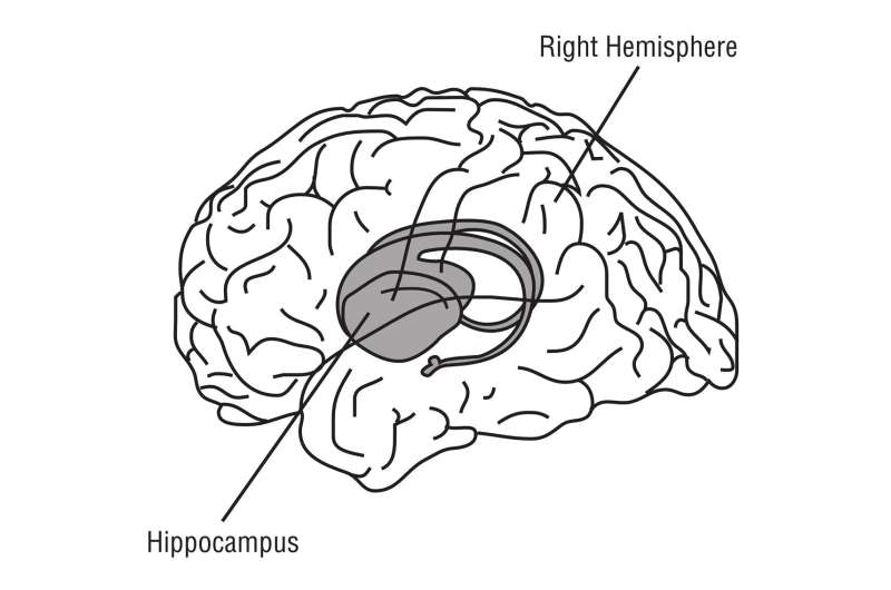
People love stories. We find it easier to remember events when they are part of an overarching narrative. But in real life, the chapters of a story don't follow smoothly one from another. Other things happen in between. A new brain imaging study from the Center for Neuroscience at the University of California, Davis, shows that the hippocampus is the brain's storyteller, connecting separate, distant events into a single narrative. The work is published Sept. 29 in Current Biology.


30 Oct 2012--Cohn-Sheehy and colleagues at Professor Charan Ranganath's Dynamic Memory Laboratory at the Center for Neuroscience used functional MRI to image the hippocampus of volunteers as they learned and recalled a series of short stories."Things that happen in real life don't always connect directly, but we can remember the details of each event better if they form a coherent narrative," said Brendan Cohn-Sheehy, a M.D./Ph.D. student at UC Davis and first author on the paper.

The stories, created specifically for the study, featured main and side characters and an event. The stories were constructed so that some formed connected, two-part narratives and others did not.

The researchers played recordings of the stories to the volunteers in the fMRI scanner. The next day, they scanned them again as the volunteers recalled the stories. The researchers compared the patterns of activity in the hippocampus between learning and recalling the different stories.

As expected, they saw more similarity for learning pieces of a coherent story than for stories that did not connect. The results show the coherent memories being woven together, Cohn-Sheehy said.

"When you get to the second event, you're reaching back to the first event and embedding part of it in the new memory," he said.

Hippocampus weaves memories

Next, they compared hippocampal patterns during learning and retrieval. They found that when recalling stories that formed a coherent narrative, the hippocampus activates more information about the second event than when recalling non-connected stories.

"The second event is where the hippocampus is forming a connected memory," Cohn-Sheehy said.

When the researchers tested the volunteers' memory of stories, they found that the ability to bring back hippocampal activity of the second event was linked to the amount of detail the volunteers could recall.

While other parts of the brain are involved in the process of memory, the hippocampus appears to bring pieces together across time and form them into connected, narrative memories, Cohn-Sheehy said.

The work is part of a new era in memory research. Traditionally, in neuroscience, researchers have studied the basic processes of memory involving disconnected pieces of information, whereas psychology has a tradition of studying how memory works to capture and connect events in the "real world." These two camps are starting to merge, Cohn-Sheehy said.

"We're using brain imaging to get at realistic memory processes," he said.

Research on memory processes could ultimately lead to better clinical tests for early stages of memory decline in aging or dementia, or for assessing damage to memory from brain injuries.

Additional authors on the study are: Jordan Crivelli-Decker, Kamin Kim and Alexander Barnett at UC Davis; and Angelique Delarazan, Zachariah Reagh and Jeffrey Zacks at Washington University St. Louis. The work was partly funded by the U.S. Office of Naval Research and the National Institute of Aging.


More information: The hippocampus constructs narrative memories across distant events, Current Biology (2021). DOI: 10.1016/j.cub.2021.09.013
Journal information: Current Biology 

Sunday, September 26, 2021


Four keys to healthy aging

exercise
Credit: Unsplash/CC0 Public Domain

The "secrets" to healthy aging are anything but secret. They're the conventional medical recommendations most people will hear from their doctor at a typical checkup: exercise, eat a healthy diet and keep up-to-date with your doctors' visits and vaccinations.

26 sep 2021--Each year, more evidence emerges to help us determine what that healthy diet should look like and how much exercise is enough to age better and live longer.

"You need to do more exercise than you think," says Dr. Jorge Camilo Mora, director of geriatric medical education at the Herbert Wertheim College of Medicine.

Thirty minutes of exercise five days a week has traditionally been the recommendation for adults. But Mora said many people don't realize their daily walks are probably not enough.

"Exercise has to be more intense than a walk," he said. "Ideally, it would combine flexibility, strength, and balance."

A moderate-intensity, brisk 30-minute walk five days a week, plus a muscle-strengthening workout twice a week meets the Centers for Disease Control's recommendations, for example.

For diet, Mora recommends moving toward more plant-based meals. That is, getting more of your daily nutrition from fruits and vegetables and less of it from meat.

Adopting a plant-based diet is associated with a reduced risk of type 2 diabetes, cancer, heart disease and Alzheimer's disease, according to a 2020 article published in the Journal of the American College of Nutrition.

Mora notes that people who live in "Blue Zones," the parts of the world where people live longest, tend to eat diets rich in plant-based foods. These people have a much higher-than-average number of centenarians—people who live to 100 years or more. Why not borrow a bit of their wisdom and incorporate more vegetarian meals into your diet?

As we observe Healthy Aging Month this September, here are Mora's tips:

  • Exercise! At least 150 minutes per week of moderate-intensity activity. Try to find activities that will also work on your strength and flexibility.
  • Eat more plant-based foods rich in antioxidants and vitamins.
  • Get your COVID-19 booster shot around 6 months after your completed dose. Not only are older adults more susceptible to the novel coronavirus, but their immune response to the vaccine is less than in younger people.
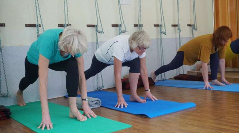
  • Get social! Social isolation is a risk factor similar to smoking in older adults. Mora's advice? Join a group exercise class—You'll meet your exercise goals AND keep up your social life.

More information: Hana Kahleova et al, Plant-Based Diets for Healthy Aging, Journal of the American College of Nutrition (2020). DOI: 10.1080/07315724.2020.1790442
Provided by Florida International University


New non-drug treatment holds promise for preventative therapies for Alzheimer's

New non-drug method holds promise for preventative therapies for Alzheimer's
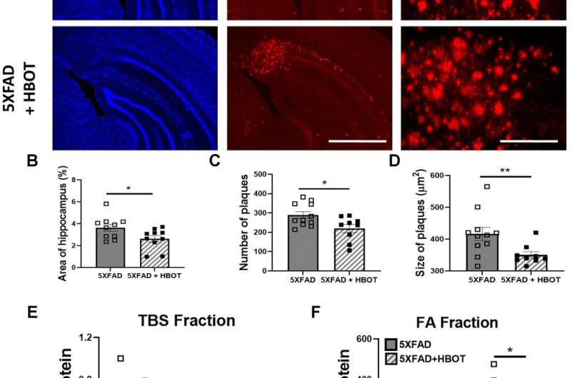
Figure 1. HBOT reduces amyloid plaques in the hippocampal area of 6-month old 5XFAD mice. Amyloid plaques were visualized by immunostaining with anti-Aβ antibodies (4G8). (A) Representative images of Aβ in the hippocampal field of HBO-treated 5XFAD (n=10, lower panel) and control 5XFAD mice (n=10, upper panel); left and middle panels, x4 magnification, scale bar: 1000 μm; right panel, x20 magnification, scale bar: 200 μm. (B) Quantification of the percentage of hippocampal area occupied by plaques. (C) Number of plaques. (D) Mean size of plaques. (E, F) Soluble Aβ was initially extracted from hippocampi with TBS by ultracentrifugation and then insoluble Aβ was extracted with 70% formic acid (FA) after ultra-centrifugation. ELISA analysis of soluble (E) and insoluble (F) Aβ40 and Aβ42 in hippocampal lysates of HBO-treated 5XFAD and control 5XFAD mice (n = 5/group). (B, C, F) -t-test, (D, F)- welch correction t-test. Values represent means ± SEM. *P < 0.05, ** P < 0.01. Credit: DOI: 10.18632/aging.203485

Approximately 50 million people worldwide live with Alzheimer's or other related forms of dementia. Alzheimer's disease leads to memory loss and impairment in cognitive function, and is the most common cause of dementia among older adults. While certain treatments can help reduce symptoms and sometimes reduce disease progression, there is currently no way to prevent or cure Alzheimer's.

26 sep 2021--Amid that backdrop, researchers from Tel Aviv University have developed a process for reversing the precursors of the disease, providing a promising foundation for new preventative therapies. This marks the first time that a non-drug therapy has proven effective in preventing the core biological processes that lead to the development of Alzheimer's, providing hope that we will now be able to fight one of the greatest challenges to the Western world.

Targeting the root of Alzheimer's

Using hyperbaric oxygen therapy (HBOT), in which subjects breathe 100% oxygen in a special chamber of high atmospheric pressure, the researchers were able to reverse brain damages associated with the biological hallmarks of Alzheimer's.

"By treating the root problem that causes cognitive deterioration with age, we are in fact mapping out the way to prevention," says co-lead researcher Prof. Shai Efrati.

Often used to treat carbon monoxide poisoning and infections that starve tissues of oxygen, hyperbaric therapy, when applied in a specific way, has previously been found capable of repairing damaged brain tissue and renewing growth of blood vessels and nerve cells in the brain. Therefore, the researchers tested its potential for Alzheimer's.

"After a series of hyperbaric treatments, elderly patients who were already suffering from memory loss showed an improvement of blood flow to the brain as well as a real improvement in cognitive performance," said co-lead investigator Prof. Uri Ashery.

The new approach devised by the researchers unequivocally improved characteristics commonly associated with Alzheimer's disease. Specifically, the hyperbaric treatment resulted in:

  • Improved memory in 16.5% of patients on average
  • Increased blood flow in 16%-23% of cases
  • Improved attention and concentration in 6% of patients
  • Improved information processing speed in 10.3% of all cases

A future without Alzheimer's?

"Our findings provide hope that we will now be able to fight one of the greatest challenges to the Western world. According to our findings, hyperbaric therapy given at a young age is likely to prevent this severe disease entirely," explains TAU team member Dr. Ronit Shapira.

The approach was first tested in laboratory settings followed by testing in patients over the age of 65 in stages of deteriorating mental function that often precede Alzheimer's and dementia. The therapy included a series of 60 treatments in hyperbaric chambers over a period of 90 days.

The study is part of a comprehensive research program focused on reversing processes of aging and its accompanying ailments. The researchers note that the findings are an encouraging step toward new approaches to preventing Alzheimer's by addressing not only the symptoms or targeting biomarkers, but the core pathology and biology responsible for the disease's development.

The findings were published in the journal Aging.


More information: Ronit Shapira et al, Hyperbaric oxygen therapy alleviates vascular dysfunction and amyloid burden in an Alzheimer's disease mouse model and in elderly patients, Aging (2021). DOI: 10.18632/aging.203485
Provided by Tel Aviv University 


Postural hypotension in older adults related to poor cognition

Postural hypotension in older adults related to poor cognition
Credit: Gerd Altmann (Pixabay)

Postural hypotension, a drastic drop in blood pressure when standing up, is linked to an increased risk of dementia and accelerated progression from cognitive impairment to dementia, even in the absence of symptoms, for instance when feeling dizzy or faint. In a study published in the journal Hypertension, researchers from Karolinska Institutet show that postural hypotension could anticipate worse cognition in old age.

26 sep 2021--Postural hypotension has already been linked with falls and cardiovascular disease. The study further showed that postural hypotension, even asymptomatic, could anticipate worse cognition in old age.

Potential implications for the public

"Older adults should actively assess their postural blood pressure to detect this condition and implement a timely intervention," says Xin Xia, Ph.D. student at the Aging Research Center, Departmant of Neurobiology, Care Sciences and Society, and co-author of the study.

This is particularly relevant since postural hypotension is usually asymptomatic, and many  with this condition may not seek medical help. The study shows that postural hypotension, even asymptomatic, appears to be harmful to cognitive function (for example memory and thinking) among older adults. This has potential implications for health care practice.

"Healthcare professionals should pay attention to the detection of postural hypotension and offer advice for timely intervention, if postural hypotension is detected," Xin Xia says.

Population-based cohort study

The study used data from from the Swedish National study on Aging and Care in Kungshomen (SNAC-K), where approximately 2500 subjects (age ≥60 years) were regularly followed. At the beginning of the follow-up, the physicians measured the participants' blood pressure when they lie down and then when they stand up after lying down for five minutes.

The drop in blood pressure was calculated when the participants stood up. The participants were then divided into two groups, one group with postural hypotension, and one without. The participants were followed for approximately 12 years, period after which the dementia risk was compared between the two groups. Later on, the subjects were divided into three groups, one group without postural hypotension, one with symptom-free postural hypotension and one with symptomatic hypotension, and subsequently their dementia risk was compared.

What happens now?

Postural hypotension in older adults may reflect poor cardiovascular health, which is what Xin Xia's research project is focused on.

"In the next step, we plan to further evaluate if ideal cardiovascular health is related to better cognitive function and a reduced risk of dementia in older adults," Xin Xia says.


More information: Xin Xia et al, From Normal Cognition to Cognitive Impairment and Dementia: Impact of Orthostatic Hypotension, Hypertension (2021). DOI: 10.1161/HYPERTENSIONAHA.121.17454
Provided by Karolinska Institutet 


Using internet in retirement boosts cognitive function

elderly computer
Credit: Unsplash/CC0 Public Domain

Using the internet during your retirement years can boost your cognitive function, a new study has found.

26 sep 2021--Researchers from Lancaster University Management School, the Norwegian University Science and Technology and Trinity College Dublin examined the cognitive function of more than 2,000 retired people from across Europe, and found that post-retirement internet usage is associated with substantially higher scores on tests.

The study, published in the Journal of Economic Behavior and Organization, uses data drawn from the Survey of Health, Aging and Retirement in Europe (SHARE) that collects information about the health, employment history and socio-economic status of older people.

Focusing on a sample of 2,105 older people from Austria, Belgium, Denmark, France, Germany, Italy, Israel, Spain, Sweden and Switzerland who have been retired since 2004, researchers examined retirees' cognitive function in both 2013 and 2015. They specifically focused on a word recall test, where individuals were asked to recall a list of 10 words immediately, and then again five minutes later.

Results found that, on average, people who used the internet after they retired were able to recall 1.22 extra words in the recall test compared to non-internet users. However, retirees who used the internet were also more likely to be male, younger, better educated, and have been retired for a shorter period. They also appear to be in better health—even though they drink and smoke more.

Dr. Vincent O'Sullivan, a co-author from Lancaster University Management School said: "Our results reveal that using the internet, post-retirement, leads to a marked reduction in the rate of cognitive decline.

"Interestingly, this protective effect was found to be most significant amongst women, with female retirees who regularly surfed the internet able to recall 2.37 more words compared to women who didn't go online. The results were also consistent among men, with retired internet users able to recall 0.94 more words than men with similar characteristics who didn't use the internet.

"We also found that retirees who used computers in their jobs before retirement were more likely to keep using computers once they retired, and hence had better cognitive function."

Researchers compared the cognitive function of retirees who used to work in jobs where computers were commonplace to retirees who worked in jobs where computers weren't often used. For example, among teachers, computers became common in the workplace much later than sectors such as financial services. Their results revealed that people with pre-retirement exposure to computers were more likely to continue to use them once they retired.

Among the overall results, the researchers also found a stark difference in the patterns of internet usage between European countries, with no more than 12% of retirees using the internet in Italy, compared to over 60% in Denmark.

"Research has shown that retirement from the workforce is a critical period for cognitive function, which declines with age and can be a predictor for a range of key health outcomes among older people," said co-author Likun Mao, formerly a Ph.D. student at Lancaster but now at Trinity College Dublin. "Although there is a widespread belief that computer usage improves older people's cognitive function—such as memory, attention, spatial abilities and problem solving—there has been mixed evidence from previous studies.

"We were able to discern that pre-retirement computer usage does not directly influence post-retirement cognitive decline, and we ensured our results referred only to post-retirement internet usage."

Professor Colin Green, of the Norwegian University Science and Technology, added: "Within our study we estimated statistical models which controlled for individuals' ages, education levels, occupational skills and years since retirement, so we are confident that our results are robust and relate only to the use of the internet, post retirement.

"This sets it apart from other studies and raises the interesting question of what it is about internet use exactly, that drives this positive effect on cognitive function. Interacting with others online, finding out information in order to attend social activities or simple tasks like shopping online can all make life easier for retirees, but we are yet to understand which, if any, of these tasks actually go as far as improving cognitive performance."

More information: Colin P. Green et al, Internet usage and the cognitive function of retirees, Journal of Economic Behavior & Organization (2021). DOI: 10.1016/j.jebo.2021.08.013
Provided by Lancaster University 


MIND diet linked to better cognitive performance

diet
Credit: CC0 Public Domain

Aging takes a toll on the body and on the mind. For example, the tissue of aging human brains sometimes develops abnormal clumps of proteins that are the hallmark of Alzheimer's disease. How can you protect your brain from these effects?

26 sep 2021--Researchers at Rush University Medical Center have found that older adults may benefit from a specific diet called the MIND diet even when they develop these protein deposits, known as amyloid plaques and tangles. Plaques and tangles are a pathology found in the brain that build up in between nerve cells and typically interfere with thinking and problem-solving skills.

Developed by the late Martha Clare Morris, ScD, who was a Rush nutritional epidemiologist, and her colleagues, the MIND diet is a hybrid of the Mediterranean and DASH (Dietary Approaches to Stop Hypertension) diets. Previous research studies have found that the MIND diet may reduce a person's risk of developing Alzheimer's disease dementia.

Now a study has shown that participants in the study who followed the MIND diet moderately later in life did not have cognition problems, according to a paper published on Sept. 14 in the Journal of Alzheimer's Disease.

"Some people have enough plaques and tangles in their brains to have a postmortem diagnosis of Alzheimer's disease, but they do not develop clinical dementia in their lifetime," said Klodian Dhana, MD, Ph.D., lead author of the paper and an assistant professor in the Division of Geriatrics and Palliative Medicine in the Department of Internal Medicine at Rush Medical College .

"Some have the ability to maintain cognitive function despite the accumulation of these pathologies in the brain, and our study suggests that the MIND diet is associated with better cognitive functions independently of brain pathologies related to Alzheimer's disease.

In this study, the researchers examined the associations of diet—from the start of the study until death—brain pathologies and cognitive functioning in older adults who participated in the Rush Alzheimer's Disease Center's ongoing Memory and Aging Project, which began in 1997 and includes people living in greater Chicago. The participants were mostly white without known dementia, and all of them agreed to undergo annual clinical evaluations while alive and brain autopsy after their death.

The researchers followed 569 participants, who were asked to complete annual evaluations and cognitive tests to see if they had developed memory and thinking problems. Beginning in 2004, participants were given an annual food frequency questionnaire about how often they ate 144 food items in previous year.

Using the questionnaire answers, the researchers gave each participant a MIND diet score based on how often the participants ate specific foods. The MIND diet has 15 dietary components, including 10 "brain-healthy food groups" and five unhealthy groups—red meat, butter and stick margarine, cheese, pastries and sweets, and fried or fast food.

To adhere to and benefit from the MIND diet, a person would need to eat at least three servings of whole grains, a green leafy vegetable and one other vegetable every day—along with a glass of wine—snack most days on nuts, have beans every other day or so, eat poultry and berries at least twice a week and fish at least once a week. A person also must limit intake of the designated unhealthy foods, limiting butter to less than 1 1/2 teaspoons a day and eating less than a serving a week of sweets and pastries, whole fat cheese, and fried or fast food.

Based on the frequency of intake reported for the healthy and unhealthy food groups, the researchers calculated the MIND diet score for each participant across the study period. An average of the MIND diet score from the start of the study until the participant's death was used in the analysis to limit measurement error. Seven sensitivity measures were calculated to confirm accuracy of the findings.

"We found that a higher MIND diet score was associated with better memory and thinking skills independently of Alzheimer's disease pathology and other common age-related brain pathologies. The diet seemed to have a protective capacity and may contribute to cognitive resilience in the elderly." Dhana said.

"Diet changes can impact cognitive functioning and risk of dementia, for better or worse," he continued. "There are fairly simple diet and lifestyle changes a person could make that may help to slow cognitive decline with aging, and contribute to brain health."

More information: Klodian Dhana et al, MIND Diet, Common Brain Pathologies, and Cognition in Community-Dwelling Older Adults, Journal of Alzheimer's Disease (2021). DOI: 10.3233/JAD-210107
Provided by Rush University Medical Center 


Important signs of blood clots in the lungs missed in patients with dementia

dementia
Credit: Pixabay/CC0 Public Domain

Patients with dementia who had signs and risk factors of a pulmonary embolism, or a blood clot in the lungs, were much less likely to be tested for pulmonary embolism than patients without dementia who had the same signs and risk factors. For example, physicians at baseline were about 1 percentage point less likely to test patients with dementia for pulmonary embolism than patients without dementia, and physicians were an additional 2.6 percentage points less likely to test patients with dementia who had an elevated heart rate—a possible sign of a pulmonary embolism—than to test those without dementia who had an elevated heart rate.

26 sep 2021--The researchers sought to determine if the presence of dementia makes a difference in how physicians evaluate patients with shortness of breath for their risk of a potentially fatal pulmonary embolism. Specifically, they examined if clinical signs and risk factors of pulmonary embolism, such as a prior history of blood clots, recent cancer, recent surgery, and elevated heart rate, were evaluated differently for patients with dementia.

The researchers studied 593,000 emergency department visits to 104 Veterans Affairs hospitals between 2011 and 2018. The sample included 7100 physicians. Patients were age 60 and older and presented with shortness of breath.

The researchers note that the study was observational and there may have been other, non-observable factors that contributed to these results. The data also did not include care wishes of patients or their caregivers, and the results may be specific to VA patients and physicians and not applicable to non-VA populations.

Physicians may miss factors associated with pulmonary embolisms, or fail to use these factors in making medical decisions, in patients who have dementia. An understanding of how physicians evaluate and make decisions in these circumstances may lead to improved care for patients with dementia.

The study is published in the peer reviewed Journal of the American Geriatrics Society.

More information: Dan P. Ly et al, How physicians evaluate patients with dementia who present with shortness of breath, Journal of the American Geriatrics Society (2021). DOI: 10.1111/jgs.17438
Provided by University of California, Los Angeles 


Intermittent fasting can help manage metabolic disease

metabolism
Credit: CC0 Public Domain

Eating your daily calories within a consistent window of 8-10 hours is a powerful strategy to prevent and manage chronic diseases such as diabetes and heart disease, according to a new manuscript published in the Endocrine Society's journal, Endocrine Reviews.

26 sep 2021--Time-restricted eating is a type of intermittent fasting that limits your food intake to a certain number of hours each day. Intermittent fasting is one of the most popular diet trends, and people are using it to lose weight, improve their health and simplify their lifestyles.

"People who are trying to lose weight and live a healthier lifestyle should pay more attention to when they eat as well as what they eat. Time-restricted eating is an easy-to-follow and effective dietary strategy that requires less mental math than counting calories," said Satchidananda Panda, Ph.D., of the Salk Institute for Biological Studies in La Jolla, Calif. "Intermittent fasting can improve sleep and a person's quality of life as well as reduce the risk of obesity, diabetes and heart disease."

In the manuscript, the researchers explore the science behind time-restricted eating, recent clinical studies and the scope for future research to better understand its health benefits. Recent research has revealed that genes, hormones and metabolism rise and fall at different times of the 24-hour day. Aligning our daily habit of when we eat with the body's internal clock can optimize health and reduce the risk or disease burden of chronic conditions like diabetes, heart disease and liver disease.

"Eating at random times breaks the synchrony of our internal program and make us prone to diseases," said Panda. "Intermittent fasting is a lifestyle that anyone can adopt. It can help eliminate health disparities and lets everyone live a healthy and fulfilling life."


More information: Emily Manoogian et al, Time-restricted eating for the prevention and management of metabolic diseases, Endocrine Reviews (2021). doi.org/10.1210/endrev/bnab027
Provided by The Endocrine Society


Pelvic floor experts issue new recommendations for magnetic resonance defecography

bowel
Credit: CC0 Public Domain

Magnetic resonance defecography (MRD) has emerged as a powerful, noninvasive imaging technique to evaluate the many possible causes of problems in passing bowel movements. A new set of consensus recommendations for performing and interpreting the results of MRD has been published by Diseases of the Colon & Rectum (DC&R), the official journal of the American Society of Colon and Rectal Surgeons (ASCRS).

26 sep 2021--The recommendations were developed by the Pelvic Floor Disorders Consortium (PFDC), a multidisciplinary organization representing the wide range of professionals involved in diagnosis and treatment of pelvic floor disorders. The new recommendations are now available on the DC&R website and appear in the journal's October issue. Liliana Bordeianou, MD, MPH, of Massachusetts General Hospital Pelvic Floor Disorders Center and Harvard Medical School was senior author of the consensus statement.

Recommendations include consensus definitions and interpretation templates for MRD

Magnetic resonance defecography is a special type of MRI scan that provides detailed, high-contrast images of the complex anatomy of the pelvic floor. Images are obtained at various stages of defecation, providing detailed information for physicians to diagnose the cause of incontinence, constipation, pelvic organ prolapse, and other conditions that interfere with the ability to pass stool. Compared to other tests, MRD provides both functional and structural information in patients with symptoms of defecatory dysfunction

Previous guidelines for performing MRD have lacked input from the many specialties—including colon and rectal surgeons, gynecologists, radiologists, urologists, and others—involved in the diagnosis and treatment of pelvic floor disorders. Differences between and among specialties may lead to variations in how the physicians interpret and use the findings of MRD. That's a special concern in the care of patients with pelvic floor disorders, who often have complex symptoms and seek care from several different professionals.

The recommendations were developed "with the explicit goal of inviting and including representatives from all relevant clinical specialties for whom MRD holds clinical significance," according to the new statement. The PFDC Working Group comprised an international roster of 24 multidisciplinary experts, who followed a formal process to develop and approve consensus recommendations in key areas.

The recommendations address important factors in performing the MRD technique, including patient positioning and the use of contrast material to provide high-quality images of the pelvic floor anatomy and function. The Working Group emphasizes the importance of education and coaching to ensure that patients understand and can follow the maneuvers needed to perform the examination.

The new document includes recommendations for interpretation of MRD scans, enabling identification and grading of any abnormalities: For example, problems with movement of the pelvic floor muscles (dyssynergia) or abnormalities interfering with stool evacuation (such as rectocele, intussusception, or rectal prolapse).

Figures and videos illustrating the MRD appearance of these findings are included in the print and online versions of the new document. The report also presents a standard template for reporting the findings and interpretation of the MRD, with the goal of promoting shared understanding and improved communication among all professionals involved in the care of patients with pelvic floor disorders. Endorsed by six national and international medical societies including ASCRS, the consensus statement is being simultaneously published in four leading specialty journals.

"The goal of this effort was to create a universal set of recommendations and language for MRD technique, interpretation, and reporting that can be utilized and carry the same significance across disciplines," Dr. Bordeianou and coauthors conclude. While the recommendations are not comprehensive, they "should be advocated as the minimum requirements when performing and interpreting MRD in patients with evacuation disorders of the pelvic floor."


More information: Brooke H. Gurland et al, Consensus Definitions and Interpretation Templates for Magnetic Resonance Imaging of Defecatory Pelvic Floor Disorders, Diseases of the Colon & Rectum (2021). DOI: 10.1097/DCR.0000000000002155