Thursday, December 17, 2020

 LED lights found to kill coronavirus

led
Credit: CC0 Public Domain

Researchers from Tel Aviv University (TAU) have proven that the coronavirus can be killed efficiently, quickly, and cheaply using ultraviolet (UV) light-emitting diodes (UV-LEDs). They believe that the UV-LED technology will soon be available for private and commercial use.

17 december 2020--This is the first study conducted on the disinfection efficiency of UV-LED irradiation at different wavelengths or frequencies on a virus from the family of coronaviruses. The study was led by Professor Hadas Mamane, Head of the Environmental Engineering Program at TAU's School of Mechnical Engineering, Iby and Aladar Fleischman Faculty of Engineering. The article was published in November 2020 issue of the Journal of Photochemistry and Photobiology B: Biology.

"The entire world is currently looking for effective solutions to disinfect the coronavirus," said Professor Mamane. "The problem is that in order to disinfect a bus, train, sports hall, or plane by chemical spraying, you need physical manpower, and in order for the spraying to be effective, you have to give the chemical time to act on the surface. Disinfection systems based on LED bulbs, however, can be installed in the ventilation system and air conditioner, for example, and sterilize the air sucked in and then emitted into the room.

"We discovered that it is quite simple to kill the coronavirus using LED bulbs that radiate ultraviolet light," she explained. "We killed the viruses using cheaper and more readily available LED bulbs, which consume little energy and do not contain mercury like regular bulbs. Our research has commercial and societal implications, given the possibility of using such LED bulbs in all areas of our lives, safely and quickly."

The researchers tested the optimal wavelength for killing the coronavirus and found that a length of 285 nanometers (nm) was almost as efficient in disinfecting the virus as a wavelength of 265 nm, requiring less than half a minute to destroy more than 99.9% of the coronaviruses. This result is significant because the cost of 285 nm LED bulbs is much lower than that of 265 nm bulbs, and the former are also more readily available.

Eventually, as the science develops, the industry will be able to make the necessary adjustments and install the bulbs in robotic systems or air conditioning, vacuum, and water systems, and thereby be able to efficiently disinfect large surfaces and spaces. Professor Mamane believes that the technology will be available for use in the near future.

It is important to note that it is very dangerous to try to use this method to disinfect surfaces inside homes. To be fully effective, a system must be designed so that a person is not directly exposed to the light.

In the future, the researchers will test their unique combination of integrated damage mechanisms and more ideas they recently developed on combined efficient direct and indirect damage to bacteria and viruses on different surfaces, air, and water.
More information: Yoram Gerchman et al, UV-LED disinfection of Coronavirus: Wavelength effect, Journal of Photochemistry and Photobiology B: Biology (2020). DOI: 10.1016/j.jphotobiol.2020.112044
Provided by Tel Aviv University 

Smartphone camera used to diagnose viral infections

Smartphone camera used to diagnose various viral infections
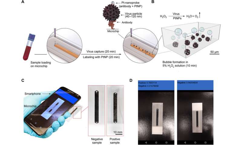
Three-dimensional schematic of the CNN–nanoparticle-enabled smartphone for virus detection. The detection process comprises three main steps. (A) Virus capture and labeling using Pt-nanoprobes. The samples are loaded into microchips modified with mAbs against the virus envelope protein and incubated to allow the capture of target virus for 20 min. The captured virus particles are then labeled with Pt-nanoprobes within 20 min. Each step of on-chip virus capture and labeling is followed by a washing step using 10 mM phosphate buffer (pH 7.4) (B) Catalyzer solution containing hydrogen peroxide (H2O2) is added to the chip and incubated for 10 min. In the presence of the captured virus, Pt-nanoprobe complex bubbles (oxygen) are formed because of the catalytic activity of PtNPs in contact with H2O2. Scale bar is set on the basis of the microchip dimensions. (C) The on-chip bubble signal is detected using the CNN–nanoparticle-enabled smartphone (NES). Photo credit: Mohamed S. Draz, Brigham and Women’s Hospital. (D) Screenshots of the results shown by the CNN-NES. Photo credit: Mohamed S. Draz, Brigham and Women’s Hospital. Credit: Science Advances (2020). DOI: 10.1126/sciadv.abd5354

A team of researchers at Brigham and Women's Hospital, Harvard Medical School, has developed a way to use a smartphone camera to test for viral infections. In their paper published in the journal Science Advances, the group describes their system, which also involves the use of an external catalytic microchip device and a smartphone system that uses a trained deep learning algorithm.

17 december 2020--As the pandemic has gripped the world for most of this year, scientists have been looking for ways to slow the spread of the next one. In this new effort, the team in Massachusetts has developed a smartphone-based system that can be used by non-medical people to test for a variety of viral infections.

The system is made up of a smartphone, an external catalytic microchip device and software. Body fluid samples are placed into a channel on the catalytic microchip device, which is then doused with a small amount of hydrogen peroxide. The resulting reaction leads to the formation of bubbles. The bubbles develop in unique patterns based in part on viruses in the fluid sample. The user points their smartphone camera at the bubbling sample and launches the deep-learning algorithm that has already been trained to identify the patterns and thereby recognize the presence of viruses. The whole process takes approximately 50 minutes. The researchers have thus far taught their system to recognize just three viruses, Zika and hepatitis B and C. But testing shows the system to be 99% accurate. They note that their system is more portable and cost effective than other solutions in the works.

The researchers suggest that their system could be rapidly trained to recognize new viruses if the need arises, and the catalytic microchip device could be sent to hot spots in the future. Such technology, the researchers suggest, could help to stop future pandemics if used widely. The researchers also note that the system could be immediately useful in infection prone areas lacking testing labs, such as third world countries.


More information: Mohamed S. Draz et al. Virus detection using nanoparticles and deep neural network–enabled smartphone system, Science Advances (2020). DOI: 10.1126/sciadv.abd5354


Sunday, December 13, 2020

Existing antihistamine drugs show effectiveness against COVID-19 virus in cell testing

Existing antihistamine drugs show effectiveness against COVID-19 virus in cell testing
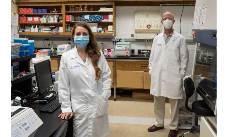
Leah Reznikov, Ph.D., and David Ostrov, Ph.D., are shown in Reznikov’s lab. Credit: UF Health/Jesse Jones

Three common antihistamine medications have been found in preliminary tests to inhibit infection of cells by the coronavirus that causes COVID-19, University of Florida Health researchers have found.

13 dec 2020--Their findings, based on laboratory tests of cells and a detailed analysis of nearly a quarter-million California patients' medical records, are published today in the journal Biochemical and Biophysical Research Communications. The data may support the launch of a randomized, controlled clinical trial to determine whether the specific antihistamines can treat or even prevent COVID-19 in humans, the researchers said.

Earlier this year, Leah Reznikov, Ph.D., an assistant professor of physiological sciences in the UF College of Veterinary Medicine and the study's principal investigator on the study, began collaborating with David A. Ostrov, Ph.D., an immunologist and associate professor in the UF College of Medicine's department of pathology, immunology and laboratory medicine. They and other colleagues set out to identify approved drugs that can interfere with the way the SARS-CoV-2 virus binds to cells. Blocking that connection essentially locks a cellular doorway that inhibits the virus's transmission to the respiratory system.

"We discovered epidemiological data showing that the usage of specific drugs was associated with a reduced likelihood of testing positive for SARS-CoV-2, the virus that causes COVID-19. We then found that these specific drugs exhibited direct antiviral activity against SARS-CoV-2 in the lab," Ostrov said.

While there is an association between the medications and infection rates, the researchers stressed there is much more to be learned and no cause and effect has been formally established.

"The fact that these drugs actually inhibit the virus in the lab does not necessarily mean that they will inhibit it actively in people—but they might," Ostrov said.

To establish their findings, the collaborators focused on angiotensin-converting enzyme-2, or ACE2, a "gateway" protein the virus uses to invade human cells. Working with a colleague at the University of California San Francisco, they analyzed medical records of nearly a quarter million California patients. People age 61 and older who had used certain antihistamines were less likely to test positive for the SARS-CoV-2 virus than those who did not take the medications, the researchers found.

Next, the researchers tested this group of antihistamines for their ability to inhibit the coronavirus in a combination of human and primate cells. Three of the drugs—hydroxyzine, diphenhydramine and azelastine—showed direct, statistically significant antiviral effects on the SARS-CoV-2 virus.

Hydroxyzine, sold as Atarax, and the nasal spray azelastine are prescription medications while diphenhydramine is sold over-the-counter as Benadryl, a treatment for cold and allergy symptoms.

The drugs were tested at different concentrations to measure how much is required to inhibit the virus.

While the findings are encouraging, Ostrov cautions against self-medicating with antihistamines as a COVID-19 prevention or treatment. So-called "off-label" use of medications should only take place after a detailed consultation with a physician, he said.

Among the three medications, azelastine was found to inhibit the SARS-CoV-2 virus at a dose that was smaller than the amount prescribed as a nasal spray. The other two antihistamines required higher drug concentrations than currently recommended dosing levels to achieve antiviral activity in cells. That doesn't make diphenhydramine any less of a potential COVID-19 therapy for now, especially considering its ubiquity and over-the-counter status, Ostrov said. Clinical trials will be necessary to establish the drugs' effectiveness in prevention, early treatment and as a secondary therapy for severe COVID-19.

Reznikov said the data suggest these three antihistamines may work by either disrupting the virus's interactions with ACE2 or by binding with another protein that may interfere with viral replication. The protein, known as a sigma receptor, is part of a cell's communications network.

Reznikov approached Ostrov in March with a hypothesis and an idea: Drugs that bind ACE2 could change disease outcomes, so she asked Ostrov to develop a list of small-molecule drug candidates.

She then pursued a strategy to screen the drugs against virus isolates of SARS-CoV-2 with Michael Norris, Ph.D., an assistant research professor of medical geography in the department of geography and the UF Emerging Pathogens Institute. In May, Reznikov was included in the UF Clinical and Translational Science Institute's Rapid-Response Translational Research Funding initiative to facilitate this project.

Although the findings are intriguing, Reznikov and Ostrov note there is still much to be learned about the mechanisms of how antihistamines interact with the SARS-CoV-2 virus.

Because of the urgency of the COVID-19 pandemic, Ostrov said there is a possibility that the antihistamine candidates could go directly to human clinical trials without first being tested in animal models. That is the case with famotidine, an antihistamine and antacid that is undergoing clinical trials elsewhere as a therapy for severely ill COVID-19 patients.


More information: Leah R. Reznikov et al. Identification of antiviral antihistamines for COVID-19 repurposing, Biochemical and Biophysical Research Communications (2020). DOI: 10.1016/j.bbrc.2020.11.095
Provided by University of Florida 

Friday, December 11, 2020

Gut microbiota plays a role in brain function and mood regulation

Gut microbiota plays a role in brain function and mood regulation
Credit: Pascal Marseaud

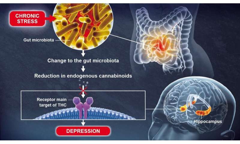
Depression is a mental disorder that affects more than 264 million people of all ages worldwide. Understanding its mechanisms is vital for the development of effective therapeutic strategies. Scientists from the Institut Pasteur, Inserm and the CNRS recently conducted a study showing that an imbalance in the gut bacterial community can cause a reduction in some metabolites, resulting in depressive-like behaviors. These findings, which show that a healthy gut microbiota contributes to normal brain function, were published in Nature Communications on December 11, 2020.

11 december 2020--The bacterial population in the gut, known as the gut microbiota, is the largest reservoir of bacteria in the body. Research has increasingly shown that the host and the gut microbiota are an excellent example of systems with mutually beneficial interactions. Recent observations also revealed a link between mood disorders and damage to the gut microbiota. This was demonstrated by a consortium of scientists from the Institut Pasteur, the CNRS and Inserm, who identified a correlation between the gut microbiota and the efficacy of fluoxetine, a molecule frequently used as an antidepressant. But some of the mechanisms governing depression, the leading cause of disability worldwide, remained unknown.

Using animal models, scientists recently discovered that a change to the gut microbiota brought about by chronic stress can lead to depressive-like behaviors, in particular by causing a reduction in lipid metabolites (small molecules resulting from metabolism) in the blood and the brain.

These lipid metabolites, known as endogenous cannabinoids (or endocannabinoids), coordinate a communication system in the body which is significantly hindered by the reduction in metabolites. Gut microbiota plays a role in brain function and mood regulation

Endocannabinoids bind to receptors that are also the main target of THC, the most widely known active component of cannabis. The scientists discovered that an absence of endocannabinoids in the hippocampus, a key brain region involved in the formation of memories and emotions, resulted in depressive-like behaviors.

The scientists obtained these results by studying the microbiotas of healthy animals and animals with mood disorders. As Pierre-Marie Lledo, Head of the Perception and Memory Unit at the Institut Pasteur (CNRS/Institut Pasteur) and joint last author of the study, explains: "Surprisingly, simply transferring the microbiota from an animal with mood disorders to an animal in good health was enough to bring about biochemical changes and confer depressive-like behaviors in the latter."

The scientists identified some bacterial species that are significantly reduced in animals with mood disorders. They then demonstrated that an oral treatment with the same bacteria restored normal levels of lipid derivatives, thereby alleviating the depressive-like behaviors. These bacteria could therefore serve as an antidepressant. Such treatments are known as "psychobiotics".

"This discovery shows the role played by the gut microbiota in normal brain function," continues Gérard Eberl, Head of the Microenvironment and Immunity Unit (Institut Pasteur/Inserm) and joint last author of the study. If there is an imbalance in the gut bacterial community, some lipids that are vital for brain function disappear, encouraging the emergence of depressive-like behaviors. In this particular case, the use of specific bacteria could be a promising method for restoring a healthy microbiota and treating mood disorders more effectively.


More information: Grégoire Chevalier et al, Effect of gut microbiota on depressive-like behaviors in mice is mediated by the endocannabinoid system, Nature Communications (2020). DOI: 10.1038/s41467-020-19931-2
Provided by Pasteur Institute 

Saturday, December 05, 2020

 How are older adults coping with the mental health effects of COVID-19?

How are older adults coping with the mental health effects of COVID-19?
McLean researchers indicate that older adults may be withstanding the mental health strains of the COVID-19 pandemic better than other age groups. Credit: McLean Hospital

Older adults are especially vulnerable to the effects of the COVID-19 pandemic—with higher risks of severe complications and death, and potentially greater difficulties accessing care and adapting to technologies such as telemedicine. A viewpoint article published in the Journal of the American Medical Association notes that there's also a concern that isolation during the pandemic could be more difficult for older individuals, which could exacerbate existing mental health conditions. Information gathered over the past several months suggests a much more nuanced picture, however.

05 december 2020--"Over the spring and summer of 2020, we were struck by a number of individual studies from all over the world that reported a consistent theme: Older adults, as a group, appeared to be withstanding the strains on mental health from the pandemic better than all other age groups," said lead author Ipsit Vahia, MD, medical director of the Geriatric Psychiatry Outpatient Services and the Institute for Technology in Psychiatryat McLean Hospital. "In this article, we highlight some of these studies and discuss resilience in older adults and what factors may be driving it."

Resilience may reflect an interaction among internal factors—such as an individual's stress response, cognitive capacity, personality traits, and physical health—and external resources like social connections and financial stability. For older adults experiencing isolation during the pandemic, having more meaningful relationships seems to be more important than having more interactions with others, and maintaining these relationships may require the use of technology to connect with loved ones.

Resilience can be supported through increased physical activity, enhanced compassion and emotional regulation, and greater social connectivity. Technology can play an important role in achieving these. "It can help maintain social connectivity, provide access to care via telemedicine, and also facilitate a range of other activities that may help cope with isolation," said Vahia. "It is increasingly becoming important for clinicians to assess patients' access and proficiency with technology as a part of care."

The authors stressed that although findings from the early months of the pandemic are encouraging and provide cause for cautious optimism, they may not reflect individual realities. "Older adults are a highly diverse group, and each person's response to the stresses of the pandemic depends on a unique set of circumstances," Vahia explained. "In addition, the current studies may not reflect specific high-risk populations with unique stressors, such as those living in underserved areas or those suffering with dementia or caregivers for people with dementia."

Importantly, the pandemic continues without a defined timeline or clear end in sight. The longer-term effects of COVID-19 on older adults' mental health, especially in countries with very high rates of disease, are unclear.


More information: Ipsit V. Vahia et al, Older Adults and the Mental Health Effects of COVID-19, JAMA (2020). DOI: 10.1001/jama.2020.21753
Provided by McLean Hospital 

Tuesday, December 01, 2020

Older adults with dementia exhibit financial 'symptoms' up to six years before diagnosis

dementia
Credit: CC0 Public Domain

A new study led by researchers at the Johns Hopkins Bloomberg School of Public Health and the Federal Reserve Board of Governors found that Medicare beneficiaries who go on to be diagnosed with dementia are more likely to miss payments on bills as early as six years before a clinical diagnosis.

01 december 2020--The study also found that beneficiaries diagnosed with dementia who had a lower educational status missed payments on bills beginning as early as seven years before a clinical diagnosis as compared to 2.5 years prior to a diagnosis for beneficiaries with higher educational status.

The study, which included researchers from the University of Michigan Medical School, also found that these missed payments and other adverse financial outcomes lead to increased risk of developing subprime credit scores starting 2.5 years before a dementia diagnosis. Subprime credit scores fall in the fair and lower range.

The findings, published online November 30 in JAMA Internal Medicine, suggest that financial symptoms such as missing payments on routine bills could be used as early predictors of dementia and highlight the benefits of earlier detection.

"Currently there are no effective treatments to delay or reverse symptoms of dementia," says lead author Lauren Hersch Nicholas, Ph.D., associate professor in the Department of Health Policy and Management at the Bloomberg School. "However, earlier screening and detection, combined with information about the risk of irreversible financial events, like foreclosure and repossession, are important to protect the financial well-being of the patient and their families."

The analysis found that the elevated risk of payment delinquency with dementia accounted for 5.2 percent of delinquencies among those six years prior to diagnosis, reaching a maximum of 17.9 percent nine months after diagnosis. Rates of elevated payment delinquency and subprime credit risk persisted for up to 3.5 years after beneficiaries received dementia diagnoses, suggesting an ongoing need for assistance managing money.

Dementia, identified as diagnostic codes for Alzheimer's Disease and related dementias in the study, is a progressive brain disorder that slowly diminishes memory and cognitive skills and limits the ability to carry out basic daily activities, including managing personal finances. About 14.7 percent of American adults over the age of 70 are diagnosed with the disease. The onset of dementia can lead to costly financial errors, irregular bill payments, and increased susceptibility to financial fraud.

For their study, the researchers linked de-identified Medicare claims and credit report data. They analyzed information on 81,364 Medicare beneficiaries living in single-person households, with 54,062 never receiving a dementia diagnosis between 1999 and 2014 and 27,302 with a dementia diagnosis during the same period. The researchers compared financial outcomes spanning 1999 to 2018 of those with and without a clinical diagnosis of dementia for up to seven years prior to a diagnosis and four years following a diagnosis. The researchers focused on missing payments for one or more credit accounts that were at least 30 days past due, and subprime credit scores, indicative of an individual's risk of defaulting on loans based on credit history.

To determine whether the financial symptoms observed were unique to dementia, the researchers also compared financial outcomes of missed payments and subprime credit scores to other health outcomes including arthritis, glaucoma, heart attacks, and hip fractures. They found no association of increased missed payments or subprime credit scores prior to a diagnosis for arthritis, glaucoma, or a hip fracture. No long-term associations were found with heart attacks.

"We don't see the same pattern with other health conditions," says Nicholas. "Dementia was the only medical condition where we saw consistent financial symptoms, especially the long period of deteriorating outcomes before clinical recognition. Our study is the first to provide large-scale quantitative evidence of the medical adage that the first place to look for dementia is in the checkbook."


More information: Lauren Hersch Nicholas et al. Financial Presentation of Alzheimer Disease and Related Dementias, JAMA Internal Medicine (2020). DOI: 10.1001/jamainternmed.2020.6432
Journal information: JAMA Internal Medicine 

Saturday, November 28, 2020

Mediterranean diet tied to 30 percent risk reduction for diabetes in Women's Health Study

mediterranean diet
Credit: CC0 Public Domain

The Mediterranean (MED) diet—rich in olive oil, fruits, vegetables, legumes, nuts and seeds—is a recommended way to reduce the risk of heart disease, type 2 diabetes and other adverse health outcomes. But exactly how and why the MED diet lowers risk for type 2 diabetes has remained unclear. In a study conducted by investigators from Brigham and Women's Hospital, researchers examined outcomes for more than 25,000 participants in the Women's Health Study, a longitudinal cohort study that followed female health professionals for more than 20 years. In a paper published in JAMA Network Open, the investigators report that women who adhered to a more MED-like diet had a 30 percent lower rate of type 2 diabetes than women who did not. The team examined several biomarkers to look for biological explanations for these results, finding key mechanisms including insulin resistance, body mass index, lipoprotein metabolism and inflammation.

28 november 2020--"Our findings support the idea that by improving their diet, people can improve their future risk of type 2 diabetes, particularly if they are overweight or have obesity," said corresponding author Samia Mora, MD, MHS, of the Brigham's divisions of Preventive Medicine and Cardiovascular Medicine and an associate professor at Harvard Medical School. "A lot of the benefit we see can be explained through just a few pathways. And it's important to note that many of these changes don't happen right away—while metabolism can change over a short period of time, our study indicates that there are longer term changes happening that may provide protection over decades."

The Women's Health Study (WHS) enrolled female health care professionals between 1992 and 1995, collecting data through December 2017. It was designed to evaluate the effects of vitamin E and low-dose aspirin on risk of heart disease and cancer. Additionally, participants were asked to complete food frequency questionnaires (FFQs) about dietary intake when the study began and answer other questions about lifestyle, medical history, demographics and more. More than 28,000 women provided blood samples at the beginning of the trial.

Mora and colleagues leveraged data from the FFQs and blood samples to investigate the relationship between the MED diet, type 2 diabetes and biomarkers that might explain the connection. To do so, they assigned each participant a MED diet intake score from 0 to 9, with points assigned for higher intake of fruits, vegetables, whole grains, legumes, nuts and fish, moderate intake of alcohol, and lower intake red meat or processed meat. The team measured a range of biomarkers, including traditional ones such as cholesterol, and more specialized ones that can only be detected using nucleic magnetic resonance. These included lipoproteins—molecules that pack and transport fats and proteins—and measures of insulin resistance, a condition in which muscle, liver and fat cells do not respond to normal amounts of insulin. Insulin resistance is often a precursor to diabetes.

Of the more than 25,000 participants in the WHS, 2,307 developed type 2 diabetes. Participants with higher MED intake at the beginning of the study (scores greater than or equal to 6) developed diabetes at rates that were 30 percent lower than participants with lower MED intake (scores less than or equal to 3). This effect was seen only among participants with a body mass index greater than 25 (overweight or obese range) and not among participants whose BMI was less than 25 (normal or underweight).

Biomarkers of insulin resistance appeared to be the biggest contributor to lower risk, followed by biomarkers of body mass index, high-density lipoprotein measures and inflammation.

"Most of this reduced risk associated with the Mediterranean diet and type 2 diabetes was explained through the biomarkers related to insulin resistance, adiposity, lipoprotein metabolism and inflammation," said first author Shafqat Ahmad, Ph.D., a researcher in the Molecular Epidemiology Unit at Uppsala University, Sweden, who helped conduct the study while working at the Brigham. "This understanding may have important downstream consequences for the primary prevention of diabetes disease."

One of the strengths of the study was its length—unlike many previous studies that have only looked at short-term effects of diets, the WHS followed participants for up to 25 years to see who developed type 2 diabetes. But the authors note several limitations, including that study participants were predominantly white and well educated, and all were female health professionals. In addition, dietary intake was self-reported and only examined at the start of the study. Biomarkers were also only measured when participants entered the study.

Mora emphasizes that insights into the biology that explains how the Mediterranean diet may help protect against diabetes could be helpful in preventive medicine and for physicians speaking to patients about dietary changes.

"Even small changes can add up over time," she said. "And there may be many biological pathways that lead to a benefit. One of the best things patients can do for future health is to improve their diet, and now we are beginning to understand why."

More information: Ahmad F et al. "Association of the Mediterranean Diet With Onset of Diabetes in the Women's Health Study" JAMA Network Open (2020). DOI: 10.1001/jamanetworkopen.2020.25466
Provided by Brigham and Women's Hospital 

Friday, November 27, 2020

 The danger of Z-drugs for dementia patients

dementia
Credit: CC0 Public Domain

Strong sleeping pills known as 'Z-drugs' are linked with an increased risk of falls, fractures and stroke among people with dementia—according to research from the University of East Anglia.

27 nov 2020--Sleep disturbance is common among people with dementia and the impact for patients and their families is significant.

To date there are no proven effective treatments available, however people with dementia are often prescribed Z-drugs (zopiclone, zaleplon, and zolpidem).

But a new study published today reveals that stronger doses of these drugs are linked with an increased risk of adverse effects.

These adverse effects were found to be similar or greater than those for higher dose benzodiazepines or 'benzos' - which are also used to treat sleep disturbance, and are known to have several adverse effects.

The team say that patients already taking higher doses of Z-drugs should not stop taking their medication suddenly, however they should seek a review with their GP.

Prof Chris Fox, from UEA's Norwich Medical School, said: "As many as 90 percent of people with dementia suffer sleep disturbances and it has a big impact on their mental and physical health, as well as that of their carers.

"Z-drugs are commonly prescribed to help people sleep—however these medicines were never licensed for dementia and they have been associated with adverse events such as falls and fracture risks in older people.

"We wanted to find out how they affect people with dementia, who are frequently prescribed them to help with sleep disturbance."

The team analysed data from 27,090 patients in England diagnosed with dementia between January 2000 and March 2016. The average age of the patients was 83 and 62 percent were women.

They looked at the adverse events for 3,532 patients who had been prescribed Z-drugs and compared them to people suffering sleep disturbance who had not been prescribed sedatives, and patients who had been prescribed benzodiazepines.

They also looked to see whether Z-drug dosage played a part in adverse outcomes.

Prof Fox said: "We studied a range of adverse outcomes including fractures, falls, deep vein thrombosis, stroke and death—over two years. And we were particularly interested to see whether higher doses led to worse outcomes."

Higher dose Z-drugs and benzodiazepines were defined as prescriptions equivalent to >7.5mg zopiclone or >5mg diazepam daily.

"For patients prescribed Z-drugs, 17 percent were given higher doses. And we found that these patients on higher doses were more at risk of falls and fractures, particularly hip fractures, and stroke—compared with patients who were not taking any medication for sleep disturbance," said Prof Fox.

Those on lower doses however (<3.75mg zopiclone or equivalent) were not found to have an increased risk of adverse outcomes.

And there were no differences in adverse events for Z-drugs compared to benzodiazepines, except lower mortality rates with Z-drugs.

Prof Fox said: "This research shows us that higher dose Z-drugs should be avoided, if possible, in people living with dementia, and non-pharmacological alternatives preferentially considered.

"Patients already taking higher dose Z-drugs should not stop taking their medication, but we recommend that they should make an appointment to see their GP for a review," he added.

Prof Clive Ballard, of the University of Exeter Medical School, who collaborated on the study, said: "Our findings serve an important caution regarding the harms of sleeping tablets in people with dementia.

"This research is a very timely and unfortunately necessary reminder that sedative medications are not a helpful way to manage social isolation during COVID-19.

"Our study also highlights the importance of research to develop non-drug approaches to help people with dementia to sleep—whether they are at home or in residential care."

Dr. Ian Maidment, Reader in Clinical Pharmacy at Aston University and lead pharmacist on the study stated: "Z-drugs are widely used to treat insomnia in people living with dementia, but are only recommended as a short-term treatment for the maximum of four weeks. Our work shows the importance of clinicians including GPs and pharmacists reviewing patients on long-term Z-drugs."


More information: 'Adverse effects of Z-drugs for sleep disturbance in people living with dementia: a population-based cohort study' is published in the journal BMC Medicine on November 24, 2020.
Provided by University of East Anglia 

Friday, November 20, 2020

 WHO Guideline Development Group advises against use of remdesivir for COVID-19

COVID-19, coronavirus
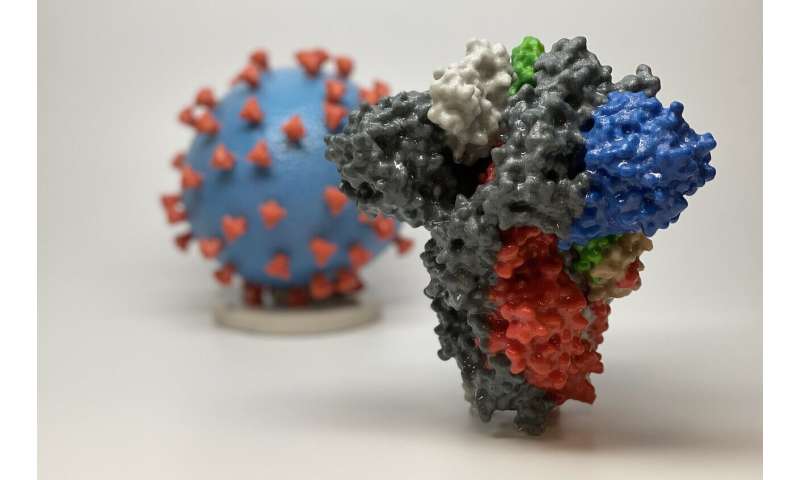
3D print of a spike protein of SARS-CoV-2, the virus that causes COVID-19--in front of a 3D print of a SARS-CoV-2 virus particle. The spike protein (foreground) enables the virus to enter and infect human cells. On the virus model, the virus surface (blue) is covered with spike proteins (red) that enable the virus to enter and infect human cells. Credit: NIH

The antiviral drug remdesivir is not suggested for patients admitted to hospital with COVID-19, regardless of how severely ill they are, because there is currently no evidence that it improves survival or the need for ventilation, say a WHO Guideline Development Group (GDG) panel of international experts in The BMJ today.

20 november 2020--The recommendation is part of a living guideline, developed by the World Health Organization with the methodological support of MAGIC Evidence Ecosystem Foundation, to provide trustworthy guidance on the management of COVID-19 and help doctors make better decisions with their patients.

Living guidelines are useful in fast moving research areas like COVID-19 because they allow researchers to update previously vetted and peer reviewed evidence summaries as new information becomes available.

Remdesivir has received worldwide attention as a potentially effective treatment for severe COVID-19 and is increasingly used to treat patients in hospital. But its role in clinical practice has remained uncertain.

Today's recommendation is based on a new evidence review comparing the effects of several drug treatments for COVID-19. It includes data from four international randomised trials involving over 7,000 patients hospitalised for COVID-19.

After thoroughly reviewing this evidence, the WHO GDG expert panel, which includes experts from around the world including four patients who have had COVID-19, concluded that remdesivir has no meaningful effect on mortality or on other important outcomes for patients, such as the need for mechanical ventilation or time to clinical improvement.

The panel acknowledged that the certainty of evidence is low and said the evidence did not prove that remdesivir has no benefit; rather, there is no evidence based on currently available data that it does improve important patient outcomes.

But given the remaining possibility of important harm, as well as the relatively high cost and resource implications associated with remdesivir (it must be given intravenously), they judged this to be an appropriate recommendation.

They also support continued enrolment into trials evaluating remdesivir, especially to provide higher certainty of evidence for specific groups of patients.

In a linked feature article, US journalist Jeremy Hsu asks what now for remdesivir, given that it is unlikely to be the lifesaving drug for the masses that many have hoped for?

The full story of remdesivir will not be known until manufacturer Gilead releases the full clinical study reports, writes Hsu, but much will depend on whether future studies are designed to test remdesivir's potential effectiveness.

In the meantime, he says alternative treatments, such as the well known, cheap, and widely available corticosteroid dexamethasone, that has been proved to reduce mortality among severely ill COVID-19 patients, are now impacting discussions about remdesivir's cost-effectiveness.


More information: François Lamontagne et al, A living WHO guideline on drugs for covid-19, BMJ (2020). DOI: 10.1136/bmj.m3379
Journal information: British Medical Journal (BMJ) 


Tuesday, November 17, 2020

 Future doctors need more nutrition education

nutrition
Credit: Pixabay/CC0 Public Domain

Medical students need more support to increase their nutrition knowledge and skills before they graduate a Griffith University study has found.

17 november 2020--The five-year study published in the journal BMJ Nutrition, Prevention and Health, examined over 100 New Zealand medical students' self-reported nutrition knowledge at three time points during their medical training by asking them to complete the same survey at the end of year 2, year 4 and year 5 of their degree.

"While the students modestly increased their nutrition knowledge and  between year 2 and year 4, the overall results were still very low,'' says lead author Associate Professor Lauren Ball from Menzies Health Institute Queensland.

"There was a clear desire for more nutrition education from all students. A key strategy to support healthy eating is to incorporate nutrition into healthcare service and doctors are expected to provide nutrition advice to patients to improve their diet when necessary. But to provide nutrition care, doctors require adequate nutrition knowledge, skills and attitudes to support the integration of such care into routine practice with patients."

Associate Professor Ball said it was recognized that graduating medical students lack nutrition knowledge and skills to actively support dietary behavior change in patients.

"This is the first study to describe how medical students' self-perceived nutrition competence changes over time during medical training. These results show that further supporting medical students to increase their knowledge and skills in this area is essential. While the study was conducted in New Zealand, Australian students face the same challenges as their counterparts across the ditch because medical programs are accredited by the same body, with identical mandatory components. Given the recognized link between nutrition and health, one approach to consider is making nutrition education compulsory for medical training."


More information: Jennifer Crowley et al. How does self-perceived nutrition competence change over time during medical training? A prospective longitudinal observational study of New Zealand medical students, BMJ Nutrition, Prevention & Health (2020). DOI: 10.1136/bmjnph-2020-000080
Provided by Griffith University 

Monday, November 16, 2020

 Link between Alzheimer's disease and gut microbiota is confirmed

alzheimers
Credit: Unsplash/CC0 Public Domain

Alzheimer's disease is the most common cause of dementia. Still incurable, it directly affects nearly one million people in Europe, and indirectly millions of family members as well as society as a whole. In recent years, the scientific community has suspected that the gut microbiota plays a role in the development of the disease.

16 november 2020--A team from the University of Geneva (UNIGE) and the University Hospitals of Geneva (HUG) in Switzerland, together with Italian colleagues from the National Research and Care Center for Alzheimer's and Psychiatric Diseases Fatebenefratelli in Brescia, University of Naples and the IRCCS SDN Research Center in Naples, confirm the correlation, in humans, between an imbalance in the gut microbiota and the development of amyloid plaques in the brain, which are at the origin of the neurodegenerative disorders characteristic of Alzheimer's disease. Proteins produced by certain intestinal bacteria, identified in the blood of patients, could indeed modify the interaction between the immune and the nervous systems and trigger the disease. These results, published in the Journal of Alzheimer's Disease, make it possible to envisage new preventive strategies based on the modulation of the microbiota of people at risk.

The research laboratory of neurologist Giovanni Frisoni, director of the HUG Memory Centre and professor at the Department of Rehabilitation and Geriatrics of the UNIGE Faculty of Medicine, has been working for several years now on the potential influence of the gut microbiota on the brain, and more particularly on neurodegenerative diseases. "We have already shown that the gut microbiota composition in patients with Alzheimer's disease was altered, compared to people who do not suffer from such disorders," he explains. "Their microbiota has indeed a reduced microbial diversity, with an over-representation of certain bacteria and a strong decrease in other microbes. Furthermore, we have also discovered an association between an inflammatory phenomenon detected in the blood, certain intestinal bacteria and Alzheimer's disease; hence the hypothesis that we wanted to test here: could inflammation in the blood be a mediator between the microbiota and the brain?"

The brain under influence

Intestinal bacteria can influence the functioning of the brain and promote neurodegeneration through several pathways: they can indeed influence the regulation of the immune system and, consequently, can modify the interaction between the immune system and the nervous system. Lipopolysaccharides, a protein located on the membrane of bacteria with pro-inflammatory properties, have been found in amyloid plaques and around vessels in the brains of people with Alzheimer's disease. In addition, the intestinal microbiota produces metabolites—in particular some short-chain fatty acids—which, having neuroprotective and anti-inflammatory properties, directly or indirectly affect brain function.

"To determine whether inflammation mediators and bacterial metabolites constitute a link between the gut microbiota and amyloid pathology in Alzheimer's disease, we studied a cohort of 89 people between 65 and 85 years of age. Some suffered from Alzheimer's disease or other neurodegenerative diseases causing similar memory problems, while others did not have any memory problems," reports Moira Marizzoni, a researcher at the Fatebenefratelli Center in Brescia and first author of this work. "Using PET imaging, we measured their amyloid deposition and then quantified the presence in their blood of various inflammation markers and proteins produced by intestinal bacteria, such as lipopolysaccharides and short-chain fatty acids."

A very clear correlation

"Our results are indisputable: certain bacterial products of the intestinal microbiota are correlated with the quantity of amyloid plaques in the brain," explains Moira Marizzoni. "Indeed, high blood levels of lipopolysaccharides and certain short-chain fatty acids (acetate and valerate) were associated with both large amyloid deposits in the brain. Conversely, high levels of another short-chain fatty acid, butyrate, were associated with less amyloid pathology."

This work thus provides proof of an association between certain proteins of the gut microbiota and cerebral amyloidosis through a blood inflammatory phenomenon. Scientists will now work to identify specific bacteria, or a group of bacteria, involved in this phenomenon.

A strategy based on prevention

This discovery paves the way for potentially highly innovative protective strategies—through the administration of a bacterial cocktail, for example, or of pre-biotics to feed the "good" bacteria in our intestine. "However, we shouldn't be too quick to rejoice," says Frisoni. "Indeed, we must first identify the strains of the cocktail. Then, a neuroprotective effect could only be effective at a very early stage of the disease, with a view to prevention rather than therapy. However, early diagnosis is still one of the main challenges in the management of neurodegenerative diseases, as protocols must be developed to identify high-risk individuals and treat them well before the appearance of detectable symptoms."


More information: Moira Marizzoni et al. Short-Chain Fatty Acids and Lipopolysaccharide as Mediators Between Gut Dysbiosis and Amyloid Pathology in Alzheimer's Disease, Journal of Alzheimer's Disease (2020). DOI: 10.3233/JAD-200306
Journal information: Journal of Alzheimer's Disease 

Provided by University of Geneva 

Friday, November 13, 2020


Common SARS-CoV-2 mutation may make coronavirus more susceptible to a vaccine

covid-19
Credit: Unsplash/CC0 Public Domain

A new study published in Science confirms that SARS-CoV-2 has mutated in a way that's enabled it to spread quickly around the world, but the spike mutation may also make the virus more susceptible to a vaccine.

13 november 2020--The new strain of coronavirus, called D614G, emerged in Europe and has become the most common in the world. Research at the University of North Carolina at Chapel Hill and the University of Wisconsin-Madison shows the D614G strain replicates faster and is more transmissible than the virus, originating in China, that spread in the beginning of the pandemic.

There were bright spots in the study findings: While the D614G strain spreads faster, in animal studies it was not associated with more severe disease, and the strain is slightly more sensitive to neutralization by antibody drugs.

The study published Nov. 12 provides some of the first concrete findings about how SARS-CoV-2 is evolving.

"The D614G virus outcompetes and outgrows the ancestral strain by about 10-fold and replicates extremely efficiently in primary nasal epithelial cells, which are a potentially important site for person-to-person transmission," said Ralph Baric, professor of epidemiology at the UNC-Chapel Hill Gillings School of Global Public Health and professor of microbiology and immunology at the UNC School of Medicine.

Baric has studied coronaviruses for more than three decades and was integral in the development of remdesivir, the first FDA-approved treatment for COVID-19.

Researchers believe the D614G strain of coronavirus dominates because it increases the spike protein's ability to open cells for the virus to enter. These crown-like spikes give the coronavirus its name.

The D614G mutation causes a flap on the tip of one spike to pop open, allowing the virus to infect cells more efficiently but also creating a pathway to the virus' vulnerable core.

With one flap open, it's easier for antibodies—like the ones in the vaccines currently being tested—to infiltrate and disable the virus.

For the recent study, Baric Lab researchers—including first author Yixuan J. Hou—worked in collaboration with Yoshihiro Kawaoka and Peter Halfmann, both virologists on faculty at the University of Wisconsin-Madison.

"The original spike protein had a 'D' at this position, and it was replaced by a 'G,'" Kawaoka said. "Several papers had already described that this mutation makes the protein more functional and more efficient at getting into cells."

That earlier work, however, relied on a pseudotyped virus that included the receptor-binding protein but was not authentic. Using reverse genetics, Baric's team replicated a matched pair of mutant SARS-CoV-2 viruses that encoded D or G at position 614 and compared basic property analysis using cell lines, primary human respiratory cells, and mouse and hamster cells.

Kawaoka and Halfmann contributed their unique coronavirus study model, which uses hamsters. The University of Wisconsin-Madison team—including Shiho Chiba, who ran the hamster experiments—performed replication and airborne transmission studies with both the original virus and the mutated version created by Baric and Hou.

They found that the mutated virus not only replicates about 10 times faster—it's also much more infectious.

Hamsters were inoculated with one virus or the other. The next day, eight uninfected hamsters were placed into cages next to infected hamsters. There was a divider between them so they could not touch, but air could pass between the cages.

Researchers began looking for replication of the virus in the uninfected animals on day two. Both viruses passed between animals via airborne transmission, but the timing was different.

With the mutant virus, the researchers saw transmission to six out of eight hamsters within two days, and to all the hamsters by day four. With the original virus, they saw no transmission on day two, though all of the exposed animals were infected by day four.

"We saw that the mutant virus transmits better airborne than the [original] virus, which may explain why this virus dominated in humans," Kawaoka said.

The researchers also examined the pathology of the two coronavirus strains. Once hamsters were infected, they presented essentially the same viral load and symptoms. (The hamsters with the mutated strain lost slightly more weight while sick.) This suggests that while the mutant virus is much better at infecting hosts, it doesn't cause significantly worse illness.

Researchers caution that the pathology results may not hold true in human studies.

"SARS-CoV-2 is an entirely new human pathogen and its evolution in human populations is hard to predict," Baric said. "New variants are continually emerging, like the recently discovered mink SARS-CoV-2 cluster 5 variant in Denmark that also encodes D614G.

"To maximally protect public health, we must continue to track and understand the consequences of these new mutations on disease severity, transmission, host range and vulnerability to vaccine-induced immunity."


More information: Yixuan J. Hou et al, SARS-CoV-2 D614G variant exhibits efficient replication ex vivo and transmission in vivo, Science  12 Nov 2020:eabe8499. DOI: 10.1126/science.abe8499
Journal information: Science