Tuesday, May 31, 2022


Cognitive impairment from severe COVID-19 equivalent to 20 years of aging, study finds
patient
Credit: Pixabay/CC0 Public Domain

Cognitive impairment as a result of severe COVID-19 is similar to that sustained between 50 and 70 years of age and is the equivalent to losing 10 IQ points, say a team of scientists from the University of Cambridge and Imperial College London.

31 may 2022--The findings, published in the journal eClinicalMedicine, emerge from the NIHR COVID-19 BioResource. The results of the study suggest the effects are still detectable more than six months after the acute illness, and that any recovery is at best gradual.

There is growing evidence that COVID-19 can cause lasting cognitive and mental health problems, with recovered patients reporting symptoms including fatigue, 'brain fog', problems recalling words, sleep disturbances, anxiety and even post-traumatic stress disorder (PTSD) months after infection. In the UK, a study found that around one in seven individuals surveyed reported having symptoms that included cognitive difficulties 12 weeks after a positive COVID-19 test.

While even mild cases can lead to persistent cognitive symptoms, between a third and three-quarters of hospitalized patients report still suffering cognitive symptoms three to six months later.

To explore this link in greater detail, researchers analyzed data from 46 individuals who received in-hospital care, on the ward or intensive care unit, for COVID-19 at Addenbrooke's Hospital, part of Cambridge University Hospitals NHS Foundation Trust. 16 patients were put on mechanical ventilation during their stay in hospital. All the patients were admitted between March and July 2020 and were recruited to the NIHR COVID-19 BioResource.

The individuals underwent detailed computerized cognitive tests an average of six months after their acute illness using the Cognitron platform, which measures different aspects of mental faculties such as memory, attention and reasoning. Scales measuring anxiety, depression and post-traumatic stress disorder were also assessed. Their data were compared against matched controls.

This is the first time that such rigorous assessment and comparison has been carried out in relation to the after effects of severe COVID-19.

COVID-19 survivors were less accurate and with slower response times than the matched control population—and these deficits were still detectable when the patients were following up six months later. The effects were strongest for those who required mechanical ventilation. By comparing the patients to 66,008 members of the general public, the researchers estimate that the magnitude of cognitive loss is similar on average to that sustained with 20 years aging, between 50 and 70 years of age, and that this is equivalent to losing 10 IQ points.

Survivors scored particularly poorly on tasks such as verbal analogical reasoning, a finding that supports the commonly-reported problem of difficulty finding words. They also showed slower processing speeds, which aligns with previous observations post COVID-19 of decreased brain glucose consumption within the frontoparietal network of the brain, responsible for attention, complex problem-solving and working memory, among other functions.

Professor David Menon from the Division of Anaesthesia at the University of Cambridge, the study's senior author, said: "Cognitive impairment is common to a wide range of neurological disorders, including dementia, and even routine aging, but the patterns we saw—the cognitive 'fingerprint' of COVID-19—was distinct from all of these."

While it is now well established that people who have recovered from severe COVID-19 illness can have a broad spectrum of symptoms of poor mental health—depression, anxiety, post-traumatic stress, low motivation, fatigue, low mood, and disturbed sleep—the team found that acute illness severity was better at predicting the cognitive deficits.

The patients' scores and reaction times began to improve over time, but the researchers say that any recovery in cognitive faculties was at best gradual and likely to be influenced by a number of factors including illness severity and its neurological or psychological impacts.

Professor Menon added: "We followed some patients up as late as ten months after their acute infection, so were able to see a very slow improvement. While this was not statistically significant, it is at least heading in the right direction, but it is very possible that some of these individuals will never fully recover."

There are several factors that could cause the cognitive deficits, say the researchers. Direct viral infection is possible, but unlikely to be a major cause; instead, it is more likely that a combination of factors contribute, including inadequate oxygen or blood supply to the brain, blockage of large or small blood vessels due to clotting, and microscopic bleeds. However, emerging evidence suggests that the most important mechanism may be damage caused by the body's own inflammatory response and immune system.

While this study looked at hospitalized cases, the team say that even those patients not sick enough to be admitted may also have tell-tale signs of mild impairment.

Professor Adam Hampshire from the Department of Brain Sciences at Imperial College London, the study's first author, said: "Around 40,000 people have been through intensive care with COVID-19 in England alone and many more will have been very sick, but not admitted to hospital. This means there is a large number of people out there still experiencing problems with cognition many months later. We urgently need to look at what can be done to help these people."

Professor Menon and Professor Ed Bullmore from Cambridge's Department of Psychiatry are co-leading working groups as part of the COVID-19 Clinical Neuroscience Study (COVID-CNS) that aim to identify biomarkers that relate to neurological impairments as a result of COVID-19, and the neuroimaging changes that are associated with these.


More information: Adam Hampshire et al, Multivariate profile and acute-phase correlates of cognitive deficits in a COVID-19 hospitalised cohort, eClinicalMedicine (2022). DOI: 10.1016/j.eclinm.2022.101417
Provided by University of Cambridge 


How can mental health professionals open their minds to psychosis?

psychosis
Credit: Pixabay/CC0 Public Domain

Effective clinical care for patients with psychosis means understanding the "lived experience" of their delusions, say researchers at the Universities of Birmingham, York and Melbourne.

31 may 2022--By understanding the intricacies of feelings, attitudes, and experiences that are interlinked with delusions, clinicians will be better able to build trust and genuine engagement with patients.

In a new study, published today in The Lancet Psychiatry, researchers collated and reviewed first-hand accounts of the experience of delusions from patients with psychosis in mental health settings. In their review, the team used a new methodological approach informed by philosophy to combine and synthesize all the available qualitative evidence. The team then developed a new model to better understand how delusions emerge and are shaped by different contexts from multiple levels of explanation.

The authors examined the experiences of more than 370 patients, described in 24 different scientific studies and found a number of themes that were common across all the studies.

For most participants, for example, delusions were not just a symptom of illness or an irrational belief. Delusions often were the most compelling way that the person could make sense of their life, in the context of a radical alteration of reality and intense emotions. While the experience could be hostile, sparking fear or panic, individuals could also experience awe or wonder, or deep meaning in their delusions.

When reality is altered in this way, individuals can struggle to make sense of their own identity, experiencing self-doubt or a loss of control or, conversely, the feeling that they are someone special, or with a unique purpose. In the latter case, the delusional experience seemed to provide a sense of coherence, purpose and belonging at a time of intense life stress and can therefore be interpreted as temporarily adaptive or beneficial.

"These experiences are complex and nuanced," said lead author Dr. Rosa Ritunnano, consultant psychiatrist and researcher at the University of Birmingham's Institute for Mental Health and the University of Melbourne. "When we train clinicians in treating psychosis, we need to ensure they are going beyond 'correcting' a dysfunction or 'curing' a symptom. Instead, by trying to understand how the delusion is shaped by the patient's emotions, life experiences and socio-cultural contexts, we can devise treatment and support that is more relevant to the individual."

The approach has particular resonance for situations in which a patient may be detained without their consent with the aim of protecting themselves from harm. The number of patients detained in this way has been steadily rising for several years, with particular ethnic groups and those with learning disabilities being particularly impacted in this process.

"One of the main issues is that all too often, the patient's voice is lost within the process of trying to help them," says Dr. Clara Humpston, one of the senior authors on the paper. "Carers, clinicians and family members all have a challenging task in learning how to listen. That means accepting that there may not be a shared view of reality, but the patient's experience is still true and valid, and ought not to be silenced."


More information: Subjective experience and meaning of delusions in psychosis: a systematic review and qualitative evidence synthesis, The Lancet Psychiatry (2022). DOI: 10.1016/S2215-0366(22)00104-3
Provided by University of Birmingham 


Fecal transplants reverse hallmarks of aging

aging
Credit: Unsplash/CC0 Public Domain

In the search for eternal youth, poo transplants may seem like an unlikely way to reverse the aging process.

31 may 2022--However, scientists at the Quadram Institute and the University of East Anglia have provided evidence, from research in mice, that transplanting fecal microbiota from young into old mice can reverse hallmarks of aging in the gut, eyes, and brain.

In the reverse experiment, microbes from aged mice induced inflammation in the brain of young recipients and depleted a key protein required for normal vision.

These findings show that gut microbes play a role in the regulating some of the detrimental effects of aging and open up the possibility of gut microbe-based therapies to combat decline in later life.

Prof Simon Carding, from UEA's Norwich Medical School and head of the Gut Microbes and Health Research Program at the Quadram Institute, said: "This ground-breaking study provides tantalizing evidence for the direct involvement of gut microbes in aging and the functional decline of brain function and vision and offers a potential solution in the form of gut microbe replacement therapy."

It has been known for some time that the population of microbes that we carry around in our gut, collectively called the gut microbiota, is linked to health. Most diseases are associated with changes in the types and behavior of bacteria, viruses, fungi and other microbes in an individual's gut.

Some of these changes in microbiota composition happen as we age, adversely affecting metabolism and immunity, and this has been associated with age-related disorders including inflammatory bowel diseases, along with cardiovascular, autoimmune, metabolic and neurodegenerative disorders.

To better understand the effects of these changes in the microbiota in old age, scientists from the Quadram Institute transferred the gut microbes from aged mice into healthy young mice, and vice versa. They then looked at how this affected inflammatory hallmarks of aging in the gut, brain and eye, which suffer from declining function in later life.

The study, published in the journal Microbiome, found that the microbiota from old donors led to loss of integrity of the lining of the gut, allowing bacterial products to cross into the circulation, which results in triggering the immune system and inflammation in the brain and eyes.

Age-related chronic inflammation, known as inflammaging, has been associated with the activation of specific immune cells found in brain. These cells were also over-activated in the young mice who received aged microbiome transplants.

In the eye, the team also found specific proteins associated with retinal degeneration were elevated in the young mice receiving microbiota from old donors.

In old mice, these detrimental changes in the gut, eye and brain could be reversed by transplanting the gut microbiota from young mice.

In ongoing studies, the team are now working to understand how long these positive effects can last, and to identify the beneficial components of the young donor microbiota and how they impact on organs distant from the gut.

The microbiota of young mice, and the old mice who received young microbiota transplants were enriched in beneficial bacteria that have previously been associated with good health in both mice and humans.

The researchers have also analyzed the products which these bacteria produce by breaking down elements of our diet. This has uncovered significant shifts in particular lipids (fats) and vitamin metabolism, which may be linked to the changes seen in inflammatory cells in the eye and brain.

Similar pathways exist in humans, and the human gut microbiota also changes significantly in later life, but the researchers caution about extrapolating their results directly to humans until similar studies in elderly humans can be performed.

A new facility for Microbiota Replacement Therapy (MRT), also known as Fecal Microbiota Transplantation (FMT) is being built in the Quadram Institute that will facilitate such trials, as well as other trials for microbiota-related conditions.

Lead author of the study, Dr. Aimee Parker from the Quadram Institute said: "We were excited to find that by changing the gut microbiota of elderly individuals, we could rescue indicators of age-associated decline commonly seen in degenerative conditions of the eye and brain.

"Our results provide more evidence of the important links between microbes in the gut and healthy aging of tissues and organs around the body. We hope that our findings will contribute ultimately to understanding how we can manipulate our diet and our gut bacteria to maximize good health in later life."

"Fecal microbiota transfer between young and aged mice reverses hallmarks of the aging gut, eye, and brain" is published in the journal Microbiome.

More information: Aimée Parker et al, Fecal microbiota transfer between young and aged mice reverses hallmarks of the aging gut, eye, and brain, Microbiome (2022). DOI: 10.1186/s40168-022-01243-w
Provided by University of East Anglia 


Labels like 'psycho' or 'schizo' can hurt. We've workshopped alternative clinical terms

It is common to hear people use stigmatizing, discriminatory and hurtful labels such as "psycho," "schizo" or "totally bipolar." Others might minimize conditions by saying they too are "a bit OCD" because they value structure and organization.

31 may 2022--This kind of everyday use of pseudo-clinical terms can be upsetting for young people who are struggling with these conditions. Worse still, it can stop them seeking care.

Clinical terms can have the same effect. For our recent research, we worked with young patients, caregivers and clinicians to develop new mental health vocabulary that carries less stigma, but remains accurate.

Mental health labels have pros and cons

Labels can provide concise and understandable descriptions of clinical and theoretical ideas. Diagnoses enable patients and health professionals to follow evidence-based advice for effective care, because best practice guidelines are available for all labeled medical conditions.

In other words, naming a condition is the first step towards identifying the best treatment available. Labels can also help create communities of individuals who share a similar clinical description, and reassure individuals they are not alone.

On the other hand, labels can result in stigma and discrimination, poor engagement with services, increased anxiety and suicidal thoughts, and poorer mental health.

The process of posing a diagnosis, may treat an individual's strengths or their vulnerabilities as abnormalities and pathologize them.

For example, a young person's vivid imagination and artistic drive—strengths that allow them to produce wonderful artwork—might be recast as a sign of illness. Or their experience of growing up in poverty and disadvantage, could be seen as the cause of their mental illness, rather than environmental factors that may have merely contributed to it.

As such, clinicians should seek to understand a person's difficulties through a holistic, humanistic and psychological perspective, prior to giving them a label.

New terms, changing approaches

In the past decade, there have been efforts to improve naming of psychiatric disorders. Attempts to update psychiatric terms and make them more culturally appropriate and less stigmatizing have resulted in renaming schizophrenia in several countries.

Proposed terms such as Si Jue Shi Tiao (thought and perceptual dysregulation) in Hong Kong, and Johyenonbyung (attunement disorder) in South Korea, have been suggested as alternatives that carry less stigma and allow a more positive view of psychiatry.

These new terms, however, were generated by experts in the field. Consumers and clients within the mental health system have rarely been consulted, until now.

Thoughts from those 'at risk'

Currently, "ultra-high risk (for psychosis)", "at-risk mental state" and "attenuated psychosis syndrome" are used to describe young people at elevated risk of developing psychosis. But these labels can be stigmatizing and damaging for the young people who receive them.

At Orygen, new, less stigmatizing ways to describe the "risk for psychosis" concept were co-developed with young people with lived experience of mental ill-health.

During focus groups, former patients were asked how they would like their experiences to be termed if they were believed to be at risk for developing a mental illness.

This discussion resulted in them generating new terms such as "pre-diagnosis stage," "potential for developing a mental illness" and "disposition for developing a mental illness."

The terms were then presented to three groups: 46 young people identified as being at risk for psychosis and currently receiving care; 24 of their caregivers; and 52 clinicians caring for young people.

Most thought these new terms were less stigmatizing than the current ones. The new terms were still judged as informative and illustrative of young people's experiences.

Patients also told us they wanted terms like these to be fully disclosed and raised early in their care. This revealed a desire of transparency when dealing with mental ill-health and clinicians.

Names have power

Labels can, and should, be revisited when stigma becomes associated with them.

Co-designing new diagnostic labels with patients, their caregivers and clinicians is empowering for all involved. Several similar projects are underway in Italy and Japan to include a cultural perspective in renaming terms related to young people at risk of developing serious mental ill health.

We hope to integrate and use more terms generated by young people in mainstream early intervention psychiatric services. We hope this will have a meaningful impact on young people's mental health by allowing better access to care and less stigmatization.


More information: Andrea Polari et al, Patients', carers' and clinicians' attitudes towards alternative terms to describe the at-risk for psychosis state, Schizophrenia Research (2021). DOI: 10.1016/j.schres.2021.08.031
Provided by The Conversation 

Researchers use AI to prompt older adults' participation in research

older adults
Credit: CC0 Public Domain

In a new study, Florida State University researchers explore the challenges of recruiting and retaining older adults to participate in research.

31 may 2022--The study also marks the first step of a broad, interdisciplinary FSU effort to increasingly use artificial intelligence in research.

In the study, published in The Gerontologist, Associate Professor of Sociology Dawn Carr identified core "motivation clusters" among older adults for research participation. Along with her 12 FSU-based co-authors, Carr suggests that identifying those clusters—"fun seekers" and "research helpers," for example—can guide recruitment and retention strategies.

"There is a lack of representation of older adults in research that leads to findings that are skewed," Carr said. "Previous guidance on how best to encourage older adults to participate in research has been one-size-fits-all. Our research finds that older adults' motivations are varied and complex."

Carr, the new director of FSU's Claude Pepper Center, and study co-author Wally Boot, a professor in the Department of Psychology, said the lack of older adults in studies prevails throughout research and has widespread consequences. They said tailored appeals can increase the number and diversity of older adults participating in research.

"The characteristics of the people participating matter since we want to be able to generalize our findings," Boot said. "And being able to recruit large samples of older adults is crucial; without large sample sizes we can't have confidence in our results."

This is the first study stemming from a larger project funded by the National Institutes of Health (NIH). The Adherence Promotion with Person-centered Technology (APPT) project aims to understand participants' motivations and daily schedules and provide just-in-time support to help them engage in behaviors that keeps them in studies.

The goal is to develop artificial intelligence-based reminder systems that encourage older adults to participate in aging-related research.

"So much momentum and time is lost when people drop out of studies, and clinical trials can fail because people don't engage in the behaviors researchers ask them to perform," Carr said. "How can we test whether a behavioral intervention reduces the risk of cognitive impairment unless participants consistently engage in that behavior over the long term?"

Carr added, "To that end, we've already learned that there are older adults who have different clusters of motivations to participate: brain health advocates, research helpers, fun seekers and multiple-motivation enthusiasts. We found that cognitive difficulties, age, employment status and previous research participation predicted membership in these categories."

Boot said artificial-intelligence approaches help predict the types of motivational messages that might resonate and keep participants on track but also the right time to deliver those messages.

"People have habits, and we can learn routine without being obtrusive," he said. "When their adherence to the intervention begins to falter, we can detect that and provide a tailored motivational message at a time when we predict they are likely available to reengage with the study."

The study is laying the groundwork for the further use of artificial intelligence, Boot said.

"Two large clinical trials will provide a very rich dataset to further develop algorithms to help predict who may be at most risk for poor adherence and the best tailored approaches to reengage them," he said. "Ultimately, we may be able to predict and prevent lapses and dropout before they happen. This is just a first step to some very exciting possibilities."


More information: Dawn C Carr et al, Motivation to Engage in Aging Research: Are There Typologies and Predictors?, The Gerontologist (2022). DOI: 10.1093/geront/gnac035
Provided by Florida State University 


Dementia: Combination of 'feelings' and measurements suggest Alzheimer's in the early stage

Alzheimer's disease
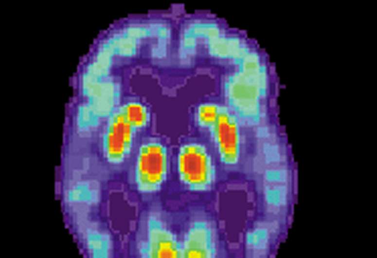
PET scan of a human brain with Alzheimer's disease. Credit: public domain

Subjective memory disorders in conjunction with conspicuous levels of beta-amyloid proteins in the cerebrospinal fluid are a strong indication of developing Alzheimer's disease. This is the conclusion of a DZNE study involving about 1,000 older adults. A team led by dementia researcher Frank Jessen reports on these findings in the journal Alzheimer's & Dementia. The study results could contribute to the early detection and treatment of Alzheimer's disease.

31 may 2022--When people feel that their memory or other mental abilities are declining, but objective tests do not reveal any deterioration, this is referred to in medicine as "subjective cognitive impairment," or SCD for short. The phenomenon has been a topic of research for several years.

"The affected individuals report cognitive problems that cause them serious concern, but which are not measurable with current techniques," explains Prof. Frank Jessen, a DZNE scientist and director of the Department of Psychiatry at University of Cologne. By now it has turned out that SCD is a risk factor, but not a conclusive warning sign for upcoming dementia. "In many individuals with SCD, there is no progressive loss of cognitive performance. To assess the individual risk more accurately, other factors have to be taken into account," the researcher says. "We have now been able to specify these. If, in addition to SCD, there is also evidence that certain proteins accumulate in the brain, then taken together that's a strong sign for a developing Alzheimer's disease."

A nationwide study

This assessment is based on a long-term DZNE study called DELCODE, which comprises ten study centers across Germany and involves several university hospitals. Within this framework, cognitive performance of almost 1,000 older women and men has been recorded annually since several years. This is done by means of established neuropsychological test procedures. In addition, the cerebrospinal fluid of many study participants is analyzed and brain volume determined by means of magnetic resonance imaging (MRI).

Jessen and his colleagues now evaluated measurement series of the individual subjects, each data set covered a period of up to five years. Mean age of the study participants was around 70 years, and they were originally recruited through memory clinics at the participating university hospitals and through newspaper advertisements. The cohort included more than 400 people with SCD at baseline and around 300 individuals who had measurable cognitive impairments—up to symptoms of dementia due to Alzheimer's disease. In addition, the cohort comprised more than 200 adults whose cognitive performance was within the normal range and who did not exhibit SCD at baseline: These "healthy" persons served as a control group. All in all, this represents one of the most comprehensive studies on SCD to date.

Biomarkers in the cerebrospinal fluid

The protein beta-amyloid, which accumulates in the brain in the course of Alzheimer's disease, played an important role in the investigations. Accumulation in the brain can be assessed indirectly—on the basis of the level of the protein in the cerebrospinal fluid: if the reading is beyond a threshold value, this is regarded as evidence that beta-amyloid is concentrating in the brain. These individuals are then considered "amyloid-positive." 83 study participants with SCD and 25 volunteers from the control group had this status. "Deposition of beta-amyloid, like SCD, is a risk factor for Alzheimer's disease. On their own, however, neither phenomenon is a clear indicator of disease. But the picture sharpens, as evidenced by our study, when these phenomena are considered together and over a longer time period," Jessen says.

Development over time

During the study period, some subjects from the SCD group and also some from the control group evolved measurable cognitive deficits. This was particularly evident in amyloid-positive subjects with SCD at baseline. In comparison, cognitive decline was much on average much lower in amyloid-positive individuals of the control group. MRI data of the brain also showed differences: The "hippocampus," a brain area divided over both brain hemispheres and considered the "control center" of memory, tended to be smaller in amyloid-positive subjects with SCD than in amyloid-positive individuals of the control group: an indication of atrophy, i.e. loss of brain mass.

Stage 2 of Alzheimer's disease

"When you add up all the findings, including the data from those subjects who already had measurable cognitive deficits at baseline, we see the combination of SCD and amyloid-positive status as a strong indicator of early-stage Alzheimer's disease," Jessen says. "If you classify Alzheimer's into six stages according to common practice, with stage 6 representing severe dementia, then, in our view, the combination of SCD and amyloid-positive status corresponds to stage 2. This occurs before the stage where measurable symptoms first appear and which is also referred to as mild cognitive impairment."

An approach for early detection

To date, there is no effective treatment for Alzheimer's disease. However, it is generally believed that therapy should begin as early as possible. "If there are measurable clinical symptoms, then the brain has already been significantly damaged. From today's perspective, treatment then has little chance of lasting success," says Jessen. "The question, therefore, is how to identify apparently healthy individuals who actually have Alzheimer's disease and are very likely to develop dementia. I consider the combination of SCD and amyloid-positive status to be a promising criterion that should be further investigated and tested in future studies."

More information: Frank Jessen et al, Subjective cognitive decline and stage 2 of Alzheimer disease in patients from memory centers, Alzheimer's & Dementia (2022). DOI: 10.1002/alz.12674
Provided by German Center for Neurodegenerative Diseases 

Frail, but living at home: Program helps elderly stay strong

Frail, but living at home: program helps elderly stay strong

It's never too late to start exercising—and the right activities might help you stay independent in your home.

31 may 2022--While building healthy habits at an earlier age can have some long-term benefits, adding physical activity can help at all ages, new research suggests.

A new study found that physically frail elderly people with low muscle mass (sarcopenia) were able to reduce their level of "mobility disability" by 22% over three years, using a program that included specific changes to their exercise habits and diets.

The formula for success involved adding extra walking, along with strength, flexibility and balance exercises, to their daily routine. It also included increasing their protein intake, according to the paper published May 11 in the BMJ.

The findings confirm the value of structured physical activity in  living on their own, said Dr. Thomas Gill, author of an editorial accompanying the study.

"We hear time and time again that the most important objective that  will report is that they want to maintain their independence as they grow older, meaning that they don't want to have to rely on others for accomplishing their day-to-day activities," said Gill, a professor of geriatric medicine at the Yale School of Medicine.

Many of the activities that people want to maintain as they grow older, from managing the distance between parking space and grocery store and then walking while shopping in the store, require a certain level of mobility, Gill noted.

"When someone loses the ability to have independent mobility, that's often a very powerful risk factor for subsequent poor outcomes, meaning that you may lose the ability to manage some more basic activities," such as bathing and dressing independently, he explained.

Study co-author Dr. Emanuele Marzetti said as a geriatrician, he is sometimes concerned about overdiagnosis and overtreatment, but not so when the prescription is exercise.

"I think we have enough evidence to prescribe physical activity and optimal nutrition without running additional trials," said Marzetti, from the Centre for Geriatric Medicine at Agostino Gemelli University Polyclinic in Rome, Italy.

Study participants included more than 1,500 men and women with an average age of 79 from 16 clinical sites in 11 European countries. About half received the exercise intervention. The others—the control group—received monthly education on healthy aging.

Participants wore an actimeter on their thighs to measure activity. All were followed for 36 months.

At the start of the trial, nearly half (47%) of those in the intervention group had mobility disability, as did 53% of the intervention group. However, all could walk about two-tenths of a mile on their own.

Physical performance scores improved more in the exercise group than in the control group after both 24 months and 36 months, researchers said.

Also, women in the group that received the intervention lost less muscle strength and less muscle mass at 24 months and less muscle mass at 36 months. No differences in muscle mass or strength were found in the men.

Guidelines do exist to indicate how to prescribe physical activity, even for frail older people, Marzetti said, but they tend not to be addressed in medical school.

"We simply took those guidelines and adapted to the conditions we were studying. We didn't do anything special. We did what should be done on a daily basis in a geriatric clinic or actually in the medical office as well," Marzetti said.

As a society, Gill said an important way to make it more possible for seniors to walk would be to remove impediments to community mobility. This would include making neighborhoods safe to walk in by having protected walking paths or indoor paths.

Gill also suggested that a more abbreviated way to identify who might be at risk of losing independent mobility, rather than conducting a full battery of tests, would be to simply evaluate gait speed.

"Ideally, these individuals could then be referred to structured physical activity programs in the community," Gill wrote in the editorial.

More information: The U.S. National Institute on Aging has more on maintaining mobility and preventing disability.

Roberto Bernabei et al, Multicomponent intervention to prevent mobility disability in frail older adults: randomised controlled trial (SPRINTT project) ,BMJ (2022)/ doi.org/10.1136/bmj-2021-068788

Journal information: British Medical Journal (BMJ) 


Sarcopenic obesity linked to dementia in elderly patients

Sarcopenic obesity linked to dementia in elderly patients, say scientists
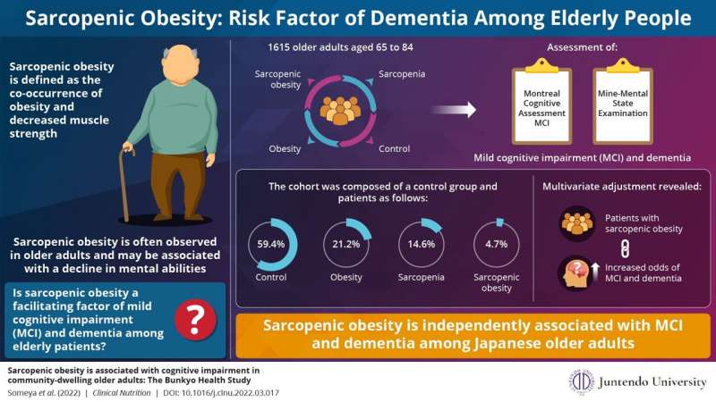
Sarcopenic obesity is independently associated with MCIand dementia among Japanese older adults. Credit: Juntendo University

Obesity, an increasingly prevalent lifestyle disease, often occurs along with poor muscle mass. This condition, called sarcopenic obesity, is evaluated based on the patients' body mass index (BMI) and handgrip strength. Interestingly, sarcopenic obesity is known to increase the risk of cognitive impairment. Dementia, a cognitive condition where memory, thinking and social abilities progressively decline, is known to significantly affect the quality of life in elderly people. Is this condition associated with sarcopenic obesity?

31 may 2022--In a new study published in Clinical Nutrition, a group of researchers led by Dr. Yoshifumi Tamura of Juntendo University, Japan, explored this very question. "If the association between sarcopenic obesity and dementia is established, appropriate preventive measures can be taken to reduce the occurrence of this condition and the risk of dementia in elderly patients," says Dr. Tamura, stressing on the importance of their study.

In the study, the researchers recruited 1,615 older Japanese adults aged 65 to 84 years participating in the Bunkyo Health Study. The researchers divided the subjects into four groups based on their sarcopenia and obesity status: those with obesity, those with sarcopenia, those with sarcopenic obesity, and those without obesity or sarcopenia (control). They studied the link between various mental processes, sarcopenia, and obesity status. Sarcopenia or poor muscle strength was determined based on a handgrip strength of less than 28 kg in men and 18 kg in women, while obesity status was given to patients with a BMI greater than 25 kg/m2. Two assessment methods were performed to establish the presence of mild cognitive impairment (MCI) and dementia. A score of less than 22 points on the Montreal cognitive assessment and less than 23 points on the Mine-Mental State Examination were used to confirm MCI and dementia respectively.

Sarcopenic obesity linked to dementia in elderly patients, say scientists
In a brand-new study, researchers from Japan have shown how co-morbidity with sarcopenia and obesity is linked with cognitive impairment in elderly Japanese people. Credit: Juntendo University

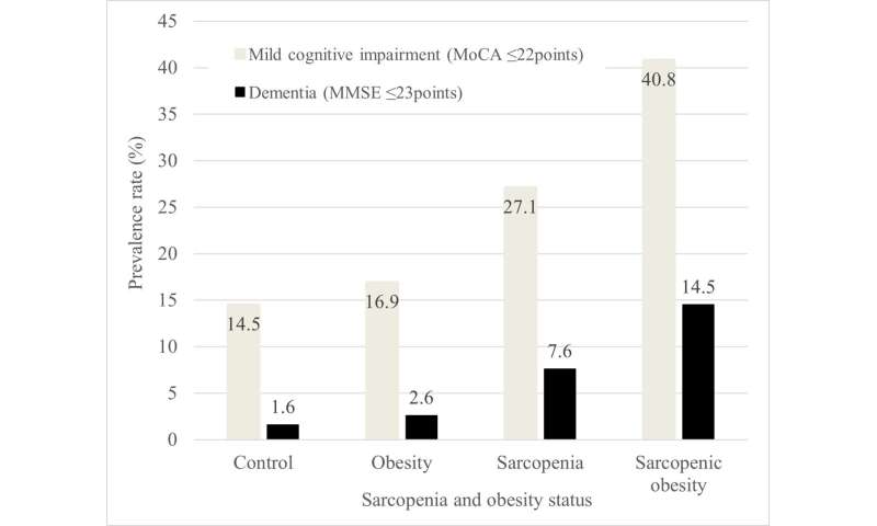
They found that 59.4% of the population had neither obesity nor sarcopenia, 21.2% had obesity, 14.6% had sarcopenia, and 4.7% of the population had sarcopenic obesity. The participants with sarcopenic obesity had the greatest rate of MCI and dementia, followed by those with sarcopenia, obesity, and finally the control group. When the team ran multivariate analyses to check for statistically relevant associations, they found that sarcopenic obesity was independently associated with an increased prevalence of MCI and dementia compared with the absence of sarcopenia and obesity. The study also showed that sarcopenia is significantly associated with dementia in women, but not in men.

"This study clearly demonstrates that sarcopenic obesity, defined by the combination of BMI and hand grip strength is associated with MCI and dementia among Japan's elderly people," says Dr. Tamura.

But what are the long-term implications of this study? Dr. Tamura says that "since we now know that there is a strong correlation between sarcopenic obesity and dementia, we may develop new treatment methods to manage the condition, thereby even reducing the prevalence of dementia."


More information: Yuki Someya et al, Sarcopenic obesity is associated with cognitive impairment in community-dwelling older adults: The Bunkyo Health Study, Clinical Nutrition (2022). DOI: 10.1016/j.clnu.2022.03.017
Provided by Juntendo University Research Promotion Center


Magnetic resonance imaging shows brain inflammation in vivo for the first time

Magnetic resonance imaging shows brain inflammation in vivo for the first time
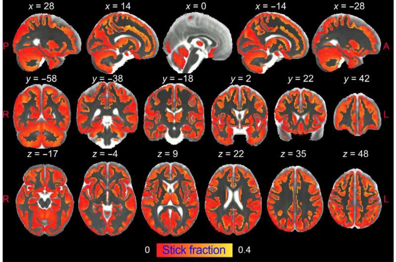
Researchers from the UMH-CSIC Neurosciences Institute have developed an innovative strategy that allows imaging of microglial and astrocyte activation in the gray matter of the brain using diffusion-weighted magnetic resonance imaging (dw-MRI). Credit: IN-CSIC-UMH

Research by Dr. Silvia de Santis and Dr. Santiago Canals, both from the Institute of Neurosciences UMH-CSIC (Alicante, Spain), has made it possible to visualize for the first time and in great detail brain inflammation using diffusion-weighted Magnetic Resonance Imaging. This detailed "X-ray" of inflammation cannot be obtained with conventional MRI, but requires data acquisition sequences and special mathematical models. Once the method was developed, the researchers were able to quantify the alterations in the morphology of the different cell populations involved in the inflammatory process in the brain.

31 may 2022--An innovative strategy developed by the researchers has made possible this important breakthrough, which is published today in the journal Science Advances and which may be crucial to change the course of the study and treatment of neurodegenerative diseases.

The research demonstrates that diffusion-weighted MRI can noninvasively and differentially detect the activation of microglia and astrocytes, two types of  cells that are at the basis of neuroinflammation and its progression.

Degenerative brain diseases such as Alzheimer's and other dementias, Parkinson's or multiple sclerosis are a pressing and difficult problem to address. Sustained activation of two types of brain cells, microglia and astrocytes, leads to chronic inflammation in the brain that is one of the causes of neurodegeneration and contributes to its progression.

However, there is a lack of non-invasive approaches capable of specifically characterizing brain inflammation in vivo. The current gold standard is positron emission tomography (PET), but it is difficult to generalize and is associated with exposure to ionizing radiation, so its use is limited in vulnerable populations and in longitudinal studies, which require the use of PET repeatedly over a period of years, as is the case in neurodegenerative diseases.

Another drawback of PET is its low spatial resolution, which makes it unsuitable for imaging small structures, with the added drawback that inflammation-specific radiotracers are expressed in multiple cell types (microglia, astrocytes and endothelium), making it impossible to differentiate between them.

In the face of these drawbacks, diffusion-weighted MRI has the unique ability to image brain microstructure in vivo noninvasively and with high resolution by capturing the random movement of water molecules in the brain parenchyma to generate contrast in MRI images.

Innovative strategy

In this study, researchers from the UMH-CSIC Neurosciences Institute have developed an innovative strategy that allows imaging of microglial and astrocyte activation in the gray matter of the brain using diffusion-weighted magnetic resonance imaging (dw-MRI).

"This is the first time it has been shown that the signal from this type of MRI (dw-MRI) can detect microglial and astrocyte activation, with specific footprints for each cell population. This strategy we have used reflects the morphological changes validated post-mortem by quantitative immunohistochemistry," the researchers note.

They have also shown that this technique is sensitive and specific for detecting inflammation with and without neurodegeneration, so that both conditions can be differentiated. In addition, it makes it possible to discriminate between inflammation and demyelination characteristic of multiple sclerosis.

Silvia de Santis says this work also demonstrates the translational value of the approach used in a cohort of healthy humans at high resolution "in which we performed a reproducibility analysis. The significant association with known microglia density patterns in the human brain supports the usefulness of the method for generating reliable glia biomarkers. We believe that characterizing, using this technique, relevant aspects of tissue microstructure during inflammation, noninvasively and longitudinally, can have a tremendous impact on our understanding of the pathophysiology of many brain conditions, and can transform current diagnostic practice and treatment monitoring strategies for neurodegenerative diseases."

To validate the model, the researchers have used an established paradigm of inflammation in rats based on intracerebral administration of lipopolysaccharide (LPS). In this paradigm, neuronal viability and morphology are preserved, while inducing, first, an activation of microglia (the brain's immune system cells), and in a delayed manner, an astrocyte response. This temporal sequence of cellular events allows glial responses to be transiently dissociated from neuronal degeneration and the signature of reactive microglia investigated independently of astrogliosis.

To isolate the imprint of astrocyte activation, the researchers repeated the experiment by pretreating the animals with an inhibitor that temporarily ablates about 90% of microglia. Subsequently using an established paradigm of neuronal damage, they tested whether the model was able to unravel neuroinflammatory "footprints" with and without concomitant neurodegeneration. "This is critical to demonstrate the utility of our approach as a platform for the discovery of biomarkers of inflammatory status in neurodegenerative diseases, where both glia activation and neuronal damage are key players," they write.

Finally, the researchers used an established paradigm of demyelination, based on focal administration of lysolecithin, to demonstrate that the biomarkers developed do not reflect the tissue alterations frequently found in brain disorders.

More information: Santiago Canals et al, Mapping microglia and astrocyte activation in vivo using diffusion MRI, Science Advances (2022). DOI: 10.1126/sciadv.abq2923www.science.org/doi/10.1126/sciadv.abq2923
Provided by Spanish National Research Council (CSIC)


Study finds health benefits of 'aging in place' at independent living facility

independent living
Credit: Unsplash/CC0 Public Domain

The American Association of Retired Persons (AARP) found the majority of older adults want to stay in their own home as they age. However, given the natural decline in health that comes with aging, some older adults may have to move into a nursing home or assisted-living facility to receive more intensive levels of care.

31 may 2022--To help older adults live independently as they "age in place," researchers at the University of Missouri analyzed eight-years of health data from 2011–2019 for more than 190 residents at TigerPlace, a senior living facility developed in partnership between the MU Sinclair School of Nursing and Americare Senior Living.

Researchers found that because registered nurse care coordinators were able to identify illnesses early and quickly in residents and provide them with appropriate care and services, most of the older adults living at TigerPlace were able to stay healthier longer, which allowed them to comfortably "age in place" and reduced their need to be transferred to a nursing home for more intensive levels of care.

TigerPlace combines the convenience and privacy of individual apartments with many recreational and socialization opportunities, such as sports bars, fitness centers, live music performances, pet therapy visits, dominoes, Bible study, bingo, volunteer opportunities and programs with local churches.

The residents at TigerPlace received health assessments from registered nurse care coordinators every six months related to cognitive functioning, completing daily tasks, depression, the risk of falling and physical functioning. Additionally, some residents chose to use noninvasive motion, bed, and depth sensors to trend level of activity, respiratory and heart rate, and fall detection. Changes in activity, new or increased falls, and assessment were used to identify illnesses, such as pneumonia or a urinary tract infection, as early as possible so interventions could be provided quickly.

"The benefits of both the regular health assessments and use of non-invasive sensors helped to keep them steady as they age comfortably," said Lori Popejoy, lead author on the study and an associate professor in the MU Sinclair School of Nursing. "The goal is to identify slight declines in health as early as possible so the right services can be put into place, whether it is connecting them with a doctor, beginning therapy or starting treatment to depression, whatever is needed based off the assessments."

Popejoy added the exercise and socialization opportunities available at TigerPlace help improve both physical and mental health outcomes, as well as reduce the risk of falls by improving muscle mass and strength. The average age of study participants was 84.

"The residents are able to use these services to enhance their quality of life in retirement, which allows them to live longer independently," Popejoy said. "For older adults that are still living at home and maybe starting to notice increased difficulty completing daily activities, or for those who are struggling with social isolation, moving to a facility like TigerPlace can be very helpful for living a healthier life longer and possibly avoiding the need to ever move to a nursing home."

The research study was interdisciplinary in nature, involving collaboration among nursing students, medical students, social workers, engineers and information technology professionals.

With May being "Older Americans Month," Popejoy has dedicated her career to improving the quality of care for older adults. She has provided hands-on clinical care in a variety of health care settings, from hospitals and nursing homes to community centers and home health care agencies.

"Longitudinal analysis of aging in place at TigerPlace: Resident function and well-being" was recently published in Geriatric Nursing.


More information: Lori Popejoy et al, Longitudinal analysis of aging in place at TigerPlace: Resident function and well-being, Geriatric Nursing (2022). DOI: 10.1016/j.gerinurse.2022.02.030
Provided by University of Missouri 


Wearable sensor finds dancers are dancing through pain

black dancers
Credit: Pixabay/CC0 Public Domain

A new Curtin University-developed wearable sensor system has tracked the movement of dancers, providing valuable insights into how they adapt to dance through disabling pain.

31 may 2022--The study, published in PLOS ONE, analyzed the movement of 52 pre-professional ballet and contemporary  students from the Western Australian Academy of Performing Arts (WAAPA). The wearable sensor system was developed as part of a collaboration between the Curtin School of Allied Health and the Curtin Institute for Computation.

Lead researcher Ph.D. student and former professional ballerina Danica Hendry, from the Curtin School of Allied Health, said that while almost all dancers experienced pain, only half of them had to stop or modify their movements.

"Fifty out of the 52 dancers said they experienced pain, with 26 reporting that this pain impacted their training/performance. Our findings indicate that the dancers are often able to continue dancing when in pain, and do so by adapting their movements, such as reducing their load," Ms. Hendry said.

"Despite a high prevalence of musculoskeletal pain, dancers' levels of pain severity and disability was generally low with the lower back and ankle/foot reported as most common."

Ms. Hendry said that existing measures, such as activity diaries and schedules, did not capture the specific movements that the dancers performed.

"Previous research exploring dancers' movement quantity has focused on measuring workload from simply looking at the daily time spent dancing," Ms. Hendry said.

"Wearable sensors have more recently been used to determine the exercise intensity of dancers during their daily training, demonstrating that while dancers participate in several hours of training per day, the majority of this time is spent doing 'low to medium intensity' exercise.

"Both approaches offer useful insights; however, until now, there was no way to provide detailed information such as the number of repetitions of movements."

Research supervisor and John Curtin Distinguished Professor Peter O'Sullivan, also from the Curtin School of Allied Health, said he was proud to have been involved with the creation of a wearable sensor system that could detect the movement quantity and quality of dancers.

"While athlete monitoring systems are commonly applied in many elite sports, it's only recently emerging within the field of dance, and only assesses the quantity of dancers' movement," Professor O'Sullivan said.

"In recent years, substantial attention has been placed on quantifying athlete training to assist in understanding the development and experience of pain and disability.

"The future application of wearable sensor technology provides the opportunity for clinicians to gain a deeper insight into the interrelationships between pain, disability, and movement in athletic populations, to better inform person-centered care.

"However, there are some challenges of the wearable sensor system that need to be addressed before more sophisticated applications can be undertaken."


More information: Danica Hendry et al, Movement quantity and quality: How do they relate to pain and disability in dancers?, PLOS ONE (2022). DOI: 10.1371/journal.pone.0268444
Journal information: PLoS ONE