Mobile applications could provide "an uninterrupted tool for crisis response" for people experiencing suicidal thoughts and behaviors, although more research is needed to establish their effectiveness, concludes a review in the March/April issue of Harvard Review of Psychiatry. The journal is published in the Lippincott portfolio by Wolters Kluwer.
10 mar 2022--In particular, apps based on an approach called ecological momentaryintervention(EMI) may offer a useful tool for managing patients at risk of suicide, according to the review by Enrique Baca-García, MD, Ph.D., of IIS-Fundación Jiménez Díaz, Madrid, Spain, and colleagues. They write, "These interventions can be useful complements to traditional care, especially in situations in which face-to-face care is not possible."
'Suicide prevention in your pocket'? So far, mixed evidence on effectiveness
Suicide remains a leading cause of potential life lost around the world, amid concerns that suicide rates may be increasing during the ongoing COVID-19 pandemic. Mobile health interventions provide an excellent opportunity to provide "low-cost, 24/7 support" for individuals at high risk of suicide, especially those with previous suicide attempts or suicidal thoughts.
Ecological momentary interventions are a particularly promising approach, with the potential to deliver as-needed help in the moment for patients experiencing suicidal thoughts and behaviors. "For instance, EMIs may allow patients to adopt coping strategies when they experience a breakdown, or to interact with the environment in different ways, such as by contacting professionals or family members during a crisis," Dr. Baca-García and coauthors write. Although EMIs have been used in other psychiatric conditions, less is known about their potential use for suicide prevention.
Dr. Baca-García and colleagues identified 27 studies of 19 different EMI interventions designed for suicide prevention. At the time of the review, 10 of the 19 interventions had at least one study evaluating effectiveness. The researchers evaluated the characteristics of the EMI interventions and the evidence for their effectiveness in suicide prevention. Eight studies, evaluating seven interventions, targeted adolescents at risk of suicide.
Safety planning was the most common component of EMI interventions. "A safety plan consists of designing a series of strategies with the support of a clinician aimed at providing support at the time of a suicidal crisis," the researchers explain. Some apps including safety plans took advantage of digital media—for example, showing pictures of loved ones, videos with relaxation techniques, or maps showing the quickest route to emergency help.
Some EMI interventions incorporated different types of approaches, such as cognitive-behavioral therapy, which teaches strategies to alleviate dysfunctional thinking or behavior; or dialectical behavior therapy, targeting healthy approaches to managing stress, emotions, and relationships.
Of the 10 EMI interventions with effectiveness studies, five had evidence of decreased suicidal thoughts and behaviors. "These mixed results suggest that there is still a long way to go before [EMI interventions] can be routinely implemented in clinical practice," Dr. Baca-García and colleagues write. Interventions based on cognitive or dialectical behavior therapy were more likely to reduce suicidal thoughts—although many of these tools also included elements of safety planning.
The studies reported high interest and good retention rates among participating patients. Adolescents and young adults may benefit most from new technologies in mental health: They are comfortable in using digital technologies and are the age group most affected by suicidal thoughts and behaviors.
"The constant advance of technology leads us to believe in the great potential for [mobile health] interventions to contribute to the field of mental health," Dr. Baca-García and co-authors conclude. "And mobile applications, with their ability to serve as an uninterrupted tool for crisis response, represent a promising field of action for suicide-prevention efforts."
More information: Laura Jiménez-Muñoz et al, Suicide Prevention in Your Pocket: A Systematic Review of Ecological Momentary Interventions for the Management of Suicidal Thoughts and Behaviors, Harvard Review of Psychiatry (2022). DOI: 10.1097/HRP.0000000000000331
Provided by Wolters Kluwer Health
Wednesday, March 09, 2022
Identification of new risk factors or early signs of Alzheimer's disease
by Institut du Cerveau (Paris Brain Institute)
Credit: Pixabay/CC0 Public Domain
What risk factors are associated with Alzheimer's up to 15 years before the onset of the first symptoms? This is a vital question for specialists of this neurodegenerative disease—which develops over many years before becoming clinically visible—who aim to improve early prevention for at-risk patients. A multidisciplinary team of researchers from the Paris Brain Institute's (INSERM/CNRS/Sorbonne University) Aramis project led by Stanley Durrleman (Inria), from INSERM/University of Bordeaux, and from Cegedim Health Data, analyzed the anonymized health records of nearly 80,000 patients consulting general practitioners in France and the United Kingdom, taken from the THIN database.
09 mar 2022--The scientists identified 10 pathologies developed more frequently by patients reporting Alzheimer's dementia within 15 years than by other patients of the same age. Their results are published in the journalThe Lancet Digital Health.
Despite the growing number of findings, our knowledge of the risk factors and early symptoms of Alzheimer's disease remains patchy and based on specific risk factor approaches. Until now, there has been no exhaustive, agnostic study conducted on a very large sample of patients that analyzes possible risk factors well ahead of the Alzheimer's diagnosis.
For the first time, a team of researchers has accessed the anonymized medical data of nearly 40,000 patients with Alzheimer's disease and of the same number of control subjects who did not develop neurodegenerative diseases over the period studied. The data was extracted from the THIN (The Health Improvement Network) database owned by Cegedim Group, an innovative technology and services company specializing in healthcare data.
The Aramis team's expertise in mathematical modeling made it possible to perform an analysis without predefined hypotheses, and test the possible link between the onset of Alzheimer's disease and 123 health factors. Statistical explorations of historical medical records yielded a list of the 10 most common conditions experienced by patients who go on to develop Alzheimer's disease within 15 years. Depression topped the list, followed by anxiety, exposure to high stress, hearing loss, constipation, cervical spondyloarthritis, memory loss, fatigue (and discomfort), and finally falls and sudden weight loss.
"The connections made allowed us to confirm known associations, such as hearing problems or depression, and other less known factors or early symptoms, such as cervical spondylosis or constipation. However, we are only reporting statistical associations. These will have to be the subject of further studies to understand the underlying mechanisms," says researcher Thomas Nedelec from the Aramis team, "The question remains as to whether the health problems encountered are risk factors, symptoms, or warning signs of the disease."
Epidemiologist and INSERM research director Carole Dufouil and neurologist Stéphane Epelbaum helped validate the methodology and interpret the relevance of these statistical associations. Although these results still need to be refined, they are already valuable for health professionals and all those involved in prevention, who could try to address these risk factors as soon as they are detected in the hopes of preventing the disease.
This work opens up several prospects, the first of which will be to expand and diversify the corpus of data studied. A grant from the European program for the study of neurodegenerative diseases (Joint Program—Neurodegenerative Disease Research) will enable the Aramis researchers to add data from Sweden and Australia to the existing pool and extend their analyses to more than 26 million data from anonymized health records. This will also enable research to be extended to other degenerative diseases (Parkinson's, Charcot's disease, multiple sclerosis, etc.).
"We hope, through this approach, to identify the common basis of these diseases and the specificities associated with each one," concludes Stanley Durrleman.
More information: Thomas Nedelec et al, Identifying health conditions associated with Alzheimer's disease up to 15 years before diagnosis: an agnostic study of French and British health records, The Lancet Digital Health (2022). DOI: 10.1016/S2589-7500(21)00275-2
Provided by Institut du Cerveau (Paris Brain Institute
Blood test for Alzheimer's proves highly accurate in large, international study
Neurologist Randall J. Bateman, MD, the Charles F. and Joanne Knight Distinguished Professor of Neurology, inspects a mass spectrometry machine at Washington University School of Medicine in St. Louis. Using mass spectrometry, Bateman and colleagues have developed a blood test that is up to 93% accurate at identifying people at risk of Alzheimer's dementia. Credit: Matt Miller/Washington University
A blood test developed at Washington University School of Medicine in St. Louis has proven highly accurate in detecting early signs of Alzheimer's disease in a study involving nearly 500 patients from across three continents, providing further evidence that the test should be considered for routine screening and diagnosis.
09 mar 2022--The study is available in the journalNeurology.
"Our study shows that the blood test provides a robust measure for detecting amyloid plaques associated with Alzheimer's disease, even among patients not yet experiencing cognitive declines," said senior author Randall J. Bateman, MD, the Charles F. and Joanne Knight Distinguished Professor of Neurology.
"A blood test for Alzheimer's provides a huge boost for Alzheimer's research and diagnosis, drastically cutting the time and cost of identifying patients for clinical trials and spurring the development of new treatment options," Bateman said. "As new drugs become available, a blood test could determine who might benefit from treatment, including those at very early stages of the disease."
Developed by Bateman and colleagues, the blood test assesses whether amyloid plaques have begun accumulating in the brain based on the ratio of the levels of the amyloid beta proteins Aβ42 and Aβ40 in the blood.
Researchers have long pursued a low-cost, easily accessible blood test for Alzheimer's as an alternative to the expensive brain scans and invasive spinal taps now used to assess the presence and progression of the disease within the brain.
Evaluating the disease using PET brain scans—still the gold standard—requires a radioactive brain scan, at an average cost of $5,000 to $8,000 per scan. Another common test, which analyzes levels of amyloid-beta and tau protein in cerebrospinal fluid, costs about $1,000 but requires a spinal tap process that some patients may be unwilling to endure.
This study estimates that prescreening with a $500 blood test could reduce by half both the cost and the time it takes to enroll patients in clinical trials that use PET scans. Screening with blood tests alone could be completed in less than six months and cut costs by tenfold or more, the study finds.
A commercial test based on Bateman's research was certified in 2020 under the Clinical Laboratory Improvement Amendments (CLIA) program. The CLIA certification program is run by the Food and Drug Administration in partnership with the Centers for Disease Control and Prevention and the Centers for Medicare and Medicaid Services.
Known as Precivity AD, the commercial version of the test is marketed by C2N Diagnostics, a Washington University startup founded by Bateman and his colleague David Holtzman, MD, the Barbara Burton and Reuben M. Morriss III Distinguished Professor of Neurology. Bateman and Holtzman are inventors on a patent the university licensed to C2N.
CLIA certification makes the test available for doctors in the United States. It is intended to provide information that will aid the medical evaluation and care of patients who already have symptoms of cognitive decline. A similar certification makes the test available in Europe. The test is not yet covered by most health insurance.
The current study shows that the blood test remains highly accurate, even when performed in different labs following different protocols, and in different cohorts across three continents.
Scientists didn't know if small differences in sampling methods, such as whether blood is collected after fasting or the type of anti-coagulant used in blood processing, could have a big impact on test accuracy because results are based on subtle shifts in amyloid beta protein levels in the blood. Differences that interfere with the precise measurement of these amyloid protein ratios could have triggered a false negative or positive result.
To confirm the test's accuracy, researchers applied it to blood samples from individuals enrolled in ongoing Alzheimer's studies in the United States, Australia and Sweden, each of which uses different protocols for the processing of blood samples and related brain imaging.
Findings from this study confirmed that the Aβ42/Aβ40 blood test using a high-precision immunoprecipitation mass spectrometry technique developed at Washington University provides highly accurate and consistent results for both cognitively impaired and unimpaired individuals across all three studies.
When blood amyloid levels were combined with another major Alzheimer's risk factor—the presence of the genetic variant APOE4—the accuracy of the blood test was 88% when compared to brain imaging and 93% when compared to spinal tap.
"These results suggest the test can be useful in identifying nonimpaired patients who may be at risk for future dementia, offering them the opportunity to get enrolled in clinical trials when early intervention has the potential to do the most good," Bateman said. "A negative test result also could help doctors rule out Alzheimer's in patients whose impairments may be related to some other health issue, disease or medication."
More information: Yan Li et al, Validation of Plasma Amyloid-β 42/40 for Detecting Alzheimer Disease Amyloid Plaques, Neurology (2021). DOI: 10.1212/WNL.0000000000013211
Provided byWashington University School of Medicine
USPSTF updates guidance on statins for primary prevention of CVD
The U.S. Preventive Services Task Force (USPSTF) recommends statins for the primary prevention of cardiovascular disease (CVD) for adults aged 40 to 75 years who have cardiovascular risk factors, with the strength of recommendation varying with cardiovascular event risk. These recommendations form the basis of a draft recommendation statement published Feb. 22 by the USPSTF.
09 mar 2022--Roger Chou, M.D., from the Oregon Health & Science University in Portland, and colleagues updated the 2016 review on statins for primary prevention in adults with cardiovascular risk. The researchers found thatstatin therapywas associated with areduced riskfor all-cause mortality, stroke,myocardial infarction, and composite cardiovascular outcomes (risk ratios, 0.92, 0.78, 0.67, and 0.72, respectively). The estimate for cardiovascular mortality was not statistically significant. The relative benefits were consistent in subgroups, including those withcardiovascular risk factorswithout marked dyslipidemia. Forolder persons, data remained sparse and imprecise. Statin therapy was not significantly associated with increased risks for serious adverse events, myalgia, liver-related harms, or diabetes.
Based on these findings, the USPSTF recommends statins for primary prevention of CVD for adults aged 40 to 75 years with one or more CVD risk factors and with an estimated 10-year risk for a cardiovascular event of 10% or greater (B recommendation). For adults aged 40 to 75 years with one or more CVD risk factors and with an estimated 10-year risk for a cardiovascular event of 7.5 to 10%, the USPSTF recommends that clinicians selectively offer statins for primary prevention (C recommendation). The evidence is currently insufficient for assessing the balance of benefits and harms of statin initiation for primary prevention of CVD and mortality in adults aged 76 years or older (I statement).
More information: The draft evidence review and recommendation statement are available for public comment from Feb. 22 to March 21, 2022.
Figure 1. Plasma and tissue osteomodulin (OMD) protein analyses in chronic kidney disease (CKD) and calcific aortic valve disease (CAVD) patients. (A) Spearman correlation between plasma OMD levels and aortic valve calcification (in Agatston scoring units) in CKD patients (n = 65). (B) OMD protein measurements in plasma from CKD patients stratified in groups according to the medial calcification grade/score (CS) of epigastric arteries from these patients (ranging from 0 to 3, where 0 signifies no arterial calcification, 1 and 2 refer to moderate calcification and 3 refers to extensive arterial calcification). Number of patients per group: n = 25 for CS = 0, n = 25 for CS = 1, n = 24 for CS = 2, n = 24 for CS = 3. One-way ANOVA multiple comparison test; data presented as mean with SD. (C) Representative histological images of epigastric arteries from CKD patients with the four different grades of calcification, immunostained for OMD (red signal) and α-SMA (green). Arrows point to OMD positive signal in the tissues. (D) Representative images from consecutive human aortic valve leaflet slides stained with Alizarin red and von Kossa to visualize calcification, or immunostained for α-SMA, OMD and RUNX2. Scale bar as indicated in all images. Insets show corresponding isotype negative control. Differences between groups were considered significant at p-values < .05 (*p < y.05). Credit: DOI: 10.1002/ctm2.682
In a new study conducted by researchers at the group Vascular Surgery, Department of Molecular Medicine and Surgery, osteomodulin was identified as a novel biomarker for vascular calcification. The results of the study were recently published in the journal Clinical and Translational Medicine.
09 mar 2022--Researchers from the Vascular surgery group, Department of Molecular Medicine and Surgery, have for a number of years studied vascularcalcificationas a prominent pathophysiological process characterized by the deposition of calcium crystals in the media or intima of vessels. In the new study led by Ph.D. candidate Nikolaos-Taxiarchis Skenteris and Assoc Prof Ljubica Matic, they utilized several large patient cohorts to investigate common mechanisms of vascular calcification in different vascular territories. Osteomodulin (OMD), a proteoglycan previously involved in bone mineralisation, was identified as a novel biomarker in this context. The study reports a consistent and broad association of both circulating and tissue OMD levels with various forms of cardiovascular calcification, highlighting its potential for further biomarker evaluation in larger clinical trials.
Calcification is a key feature of late-stage diabetes, renal and cardiovascular disease, linked to major adverse events such as myocardial infarction and stroke. Other pathological vascular changes encompass inflammation, lipid accumulation, extracellular matrix remodeling and alterations in smooth muscle cell function. Understanding mechanisms of these processes as well as their interplay in disease progression, is of key importance for finding new clinical biomarkers or therapeutic targets. This study shows that OMD is an important early modulator of cardiovascular calcification processes, enriched in association with vessel wall inflammation and osteoblastic transition of vascular smooth muscle cells, with the capacity to attenuate matrix calcification once it is secreted in the extracellular tissue.
"This study was organized as an international collaboration utilizing clinical material and data from several large cohorts of atherosclerosis (Biobank of Karolinska Endarterectomy, KI), chronic kidney disease (Kärl Tx biobank, KI in collaboration with Prof Peter Stenvinkel) and calcific aortic valve disease material obtained by Prof Leon Schurgers' group, Maastricht University. Through these collaborations we had access to unique patient material, but were also able to study advanced murine models of vascular calcification in order to understand the mechanisms regulated by OMD. Our findings imply that OMD has the potential both as a plasma biomarker and as a therapeutic target for assessing cardiovascular calcification," says Ljubica Matic, Team Leader in the Vascular Surgery group, MMK, KI and corresponding author of the study.
The study was conducted in the framework of the EU Horizon 2020 International Training Network INTRICARE which was initiated in 2017 as collaboration among Maastricht University, The Netherlands, Aachen University Clinic, Germany and Karolinska Institute. The aim of INTRICARE, that gathered totally 15 Ph.D. students across three Universities, was to study risks of vascular intimal calcification and roads to regression of cardiovascular disease.
More information: Nikolaos T. Skenteris et al, Osteomodulin attenuates smooth muscle cell osteogenic transition in vascular calcification, Clinical and Translational Medicine (2022). DOI: 10.1002/ctm2.682
Provided by Karolinska Institutet
Caregivers find remote monitoring during COVID-19 pandemic an unexpected patient safety benefit
In an opinion article appearing online Feb. 25 in the Journal of the American Medical Association, authors from University Hospitals and Case Western Reserve University write about the unexpected patient safety benefit resulting from remote monitoring of patients during the COVID-19 pandemic.
09 mar 2022--Peter Pronovost, MD, Ph.D., Melissa Cole, MSN, and Robert Hughes, DO, discuss that although COVID-19 placed excessive psychological and moral stress and work demands on patients, clinicians,health care organizationsand society, the pandemic also advanced patient safety in an unexpected way.
Prior to the pandemic, routine monitoring of patients with continuous pulse oximetry and heart rate devices was dependent on the patient's location within a hospital, usually the intensive care unit (ICU). Pulse oximeters are small electronic devices that clip onto a finger and measure the saturation of oxygen carried in red blood cells. Studies have shown that monitoring with these devices is associated with reduced death rates.
As the pandemic flooded hospitals with patients and filled ICUs, many patients received care outside of the ICU in emergency departments or general medical and surgical units. And, some medical centers advised patients with milder symptoms to stay home.
"One of the major lessons gained from the pandemic was that patients could now be monitored based on risks and needs rather than location in the hospital," said Dr. Pronovost, Chief Quality and Clinical Transformation Officer at UH and Clinical Professor of Anesthesiology and Perioperative Medicine at Case Western Reserve School of Medicine. "Home monitoring and hospital at-home models offer the potential to transform care and potentially allow a substantial proportion of hospitalized patients to receive care from home."
In their paper, the authors review benefits of remote monitoring in the hospital and at home, explore the technology advances that made it possible, describe how government payment policy changes made home monitoring sustainable, and discuss what health systems could do to launch a home monitoring program.
Ms. Cole, Vice President of Integrated Delivery Operations at UH, said that continuous remote monitoring of hospitalized patients in general medical settings not only improves outcomes, but increases accuracy.
"Technological advances have made it possible to monitor some of these patients either at home or in skilled nursing facilities. Things such as wireless monitors, cloud-based platforms and telehealth have allowed health systems to seamlessly use at-home continuous pulse oximeters to monitor patients and help them avoid hospitalizations," said Ms. Cole.
The authors write that an analysis projected remote monitoring to be potentially associated with a lower mortality rate in COVID-19 patients compared with patients without at-home monitoring: "…a mortality rate of 6 per 1,000 patients with COVID-19 compared with 26 per 1,000 patients without at-home monitoring." They write that this analysis also projected 87 percent fewer hospitalizations, 77 percent fewer deaths, and reduced per-patient costs of $11,472 compared with standard care.
"The combined use of telehealth, home health, and remote monitoring could bring some hospital-level-monitoring services to patients in their home," said Ms. Cole.
Despite these advances, however, the authors find that broad hospital and home monitoring services are not widely used by health systems. They outline several barriers that health systems must overcome.
"Health systems need to consider implementing continuous pulse oximetry and heart rate monitoring for all hospitalized patients and emergency department patients," said Dr. Hughes, Medical Director of System Operations and Logistics for UH and Assistant Professor of Emergency Medicine at Case Western Reserve School of Medicine. "With increased census, sicker patients and decreased staffing for most clinical roles, patients are at heightened risk for unrecognized deterioration. Continuous monitoring with a centralized team to monitor could improve safety and reduce workload on clinicians," he said.
Other recommendations are for health systems to create a service line to coordinate this work, to maximize value by learning how to combine and integrate these various technologies, and to create protocols for selecting and enrollment that match the patient's risks and needs with the various types of monitoring.
At University Hospitals, Ms. Cole said experiences from the pandemic helped pave the way in 2021 for UH's Hospital@Home program, the first of its kind in the health system.
"During the early days of the pandemic, we introduced UHRemote, a remote patient monitoring system that allowed us to keep tabs on COVID-19 patients' heartrate and blood oxygenation levels. A team of nurses, each with multiple screens, watched dashboards of patient data and received alerts from wearable devices if a patient's numbers were too high or too low. We learned much from the more than 2,200 patients we cared during that year, and often heard how grateful they were to not be isolated from family and alone in the hospital. We combined the experiences from UHRemote with the decades of experience of our Home Health teams, added in a few more services, and UH Hospital@Home was born," she said.
More information: Peter J. Pronovost et al, Remote Patient Monitoring During COVID-19, JAMA (2022). DOI: 10.1001/jama.2022.2040
Provided by University Hospitals Cleveland Medical Center
Study finds higher death rates in older adults with cognitive impairment during the pandemic
Findings from a new study, led by researchers at Dartmouth's Geisel School of Medicine and published in JAMA Neurology, show that higher death rates have been associated with the COVID-19 pandemic among older adults with cognitive impairment—especially in racial and ethnic minority populations and those living in nursing homes.
09 mar 2022--In 2020, COVID-19 abruptly altered the delivery of healthcare and the daily operations of nursing facilities across the U.S. These changes included a decrease in inpatient care and a transition of outpatient care to telehealth platforms. In nursing facilities, operational changes included lockdowns and strict visitation procedures, resulting in social isolation for many residents.
But the associations between those changes related to the pandemic and patient outcomes, especially in vulnerable populations, are not yet well understood.
"When you think about adult populations that are potentially most at risk for bad outcomes when things change dramatically and abruptly in healthcare, older adults with cognitive issues—like Alzheimer's disease and related dementias (ADRD)—are at or near the top of the list," says Lauren Gilstrap, MD, MPH, an assistant professor of The Dartmouth Institute for Health Policy and Clinical Practice and of medicine at Dartmouth's Geisel School of Medicine, who served as lead author on the study.
"During the pandemic, mortality rates increased across most if not all segments of society—we knew that going into the study," she continues. "Our main question was whether the increase in mortality among these more vulnerable populations was proportional or disproportional."
To find out, the researchers conducted a cross-sectional analysis using fee-for-service data on Medicare enrollees aged 65 and older. They compared mortality rates from 26.7 million enrollees in 2020 with those of 26.9 million enrollees in 2019 in four predetermined groups: people with ADRD; people without ADRD, those with ADRD living in nursing homes, and those without ADRD in nursing homes.
The investigators found that mortality was 24 percent higher among individuals with ADRD in 2020 compared to 2019, and 14 percent higher for people without ADRD. Among nursing home residents with ADRD, mortality was 36 percent higher in 2020 compared to 2019, versus 25 percent higher for those without ADRD.
The largest increases in mortality were observed among Asian, Black, and Hispanic ADRD populations.
When the researchers measured monthly COVID-19 infection rates across the country's 306 hospital referral regions (or service areas), they found that the areas with the lowest COVID-19 prevalence had no excess mortality among enrollees without ADRD.
But they found eight percent higher mortality among community-dwelling enrollees with ADRD and 14 percent higher mortality among enrollees with ADRD living in nursing homes in those same areas where there was very low COVID-19.
"That was the most interesting finding in the study," says Gilstrap. "I think what that tells us is that those deaths were probably related less to COVID and more to abrupt changes in the healthcare system and how those changes altered access to both acute and chronic care as well as community support services.
"Overall, I think our findings also highlight that as a healthcare system we really do have to think about people with cognitive limitations differently, and that more creative solutions are needed to better serve this highly vulnerable segment of society."
More information: Lauren Gilstrap et al, Trends in Mortality Rates Among Medicare Enrollees With Alzheimer Disease and Related Dementias Before and During the Early Phase of the COVID-19 Pandemic, JAMA Neurology (2022). DOI: 10.1001/jamaneurol.2022.0010
Provided by The Geisel School of Medicine at Dartmouth
30-60 mins of weekly muscle strengthening activity linked to 10-20% lower death risk
Between 30 and 60 minutes of muscle strengthening activity every week is linked to a 10-20% lower risk of death from all causes, and from cardiovascular disease, diabetes, and cancer, in particular, finds a pooled data analysis of the available evidence, published online in the British Journal of Sports Medicine.
The findings are independent ofaerobic exercise. But the analysis points to a J-shaped curve for most outcomes, with no conclusive evidence that more than an hour a week ofmusclestrengthening activity reduces the risk further still.
Physical activity guidelines recommend regular muscle strengthening activities for adults, primarily because of the known benefits for skeletal muscle health. Examples of these activities include lifting weights; working with resistance bands; push-ups, sit-ups, and squats; and heavy gardening, such as digging and shoveling.
Previous research indicates that muscle strengthening activity is associated with a lower risk of death, but it's not known what the optimal 'dose' might be.
To try and find out, the researchers scoured research databases for relevant prospective observational studies that included adults without major health issues who had been monitored for at least 2 years.
The final analysis included 16 studies out of an initial cache of 29. The earliest study was published in 2012, and most studies were carried out in the U.S., with the rest from England, Scotland, Australia, and Japan. The maximum monitoring period lasted 25 years.
Study participant numbers varied from nearly 4000 to almost 480,000, and ranged in age from 18 to 97. Twelve studies included both men and women; two included men only while three included women only. All the studies considered aerobic or other types of physical activity as well as muscle strengthening activities.
The pooled data analysis showed that muscle strengthening activities were associated with a 10–17% lower risk of death from any cause, as well as death from heart disease and stroke, cancer, diabetes, and lung cancer.
No association was found between muscle strengthening and a reduced risk of specific types of cancer, including those of the bowel, kidney, bladder or pancreas.
A J-shaped curve emerged, with a maximum risk reduction of between 10–20% at approximately 30–60 minutes/week of muscle strengthening activities for death from any cause, cardiovascular disease, and all cancer.
An L-shaped association was observed for diabetes, with a large risk reduction up to 60 minutes/week of muscle strengthening activities, after which there was a gradual tapering off.
Joint analysis of muscle strengthening and aerobic activities showed that the reduction in risk of death from any cause, cardiovascular disease, and cancer was even greater when these two types of activities were combined: 40%, 46%, and 28% lower, respectively.
The researchers acknowledge certain limitations to their findings, the main one of which was that data from only a few studies were pooled for each of the outcomes studied. The included studies also relied on subjective assessment of muscle strengthening activities.
Because most of the studies were carried out in the US, the results might not be more widely applicable, caution the researchers, who add that the included studies were all observational rather than clinical trials.
Given the J-shaped associations, the potential of a higher volume of muscle strengthening activities on the reduction in risk of death is unclear, they write.
But they conclude: "The combination of muscle strengthening and aerobic activities may provide a greater benefit for reducing all-cause, [cardiovascular disease], and total cancer mortality.
"Given that the available data are limited, further studies—such as studies focusing on a more diverse population—are needed to increase the certainty of the evidence."
More information: Muscle-strengthening activities are associated with lower risk and mortality in major non-communicable diseases: a systematic review and meta-analysis of cohort studies, British Journal of Sports Medicine (2022). DOI: 10.1136/bjsports-2021-105061
Provided by British Medical Journal
Women are more susceptible to Alzheimer's: New evidence
Epidemiological studies have shown that women are twice as likely as men to develop Alzheimer's disease (AD), but the cause of this phenomenon has been unclear.
09 mar 2022--Now, however, a study led by Prof. Keqiang Ye from the Shenzhen Institute of Advanced Technology (SIAT) of the Chinese Academy of Sciences provides a clear answer to this mystery that has puzzled mankind for decades.
Their findings were published in Nature on March 2.
Integrating their previous studies, Prof. Ye's team has established the theory that the C/EBPβ/AEP pathway is the core factor driving the pathogenesis of neurodegenerative diseases.
"Based on this theory, our team searched for female hormones that are dramatically changed during menopause and tested which hormone selectively activates the C/EBPβ/AEP pathway," said Prof. Ye.
Prof. Ye's team identified follicle-stimulating hormone (FSH) as the major pathogenic factor.
"During menopause, the serum concentration of FSH strongly increases, binding to the cognate FSH receptor on neurons and activating the C/EBPβ/AEP pathway. This results in Aβ and Tau pathologies, leading to the development of AD," said Dr. Zaidi Mone, co-corresponding author of the study and a tenured professor at the Mount Sinai School of Medicine in New York.
The researchers employed different methods to demonstrate this finding. Using ovariectomized mice, they used anti-FSH antibody treatment to block FSH and inactivate the C/EBPβ/AEP pathway. They also deleted FSH receptor (FSHR) expression in neurons to abolish the binding of FSH to FSHR in the hippocampus. Both of these methods alleviated pathology and cognitive dysfunction. In addition, knockdown of C/EBPβ in the AD mice model decreased AD pathologies.
Besides working with female mice, the researchers also injected FSH into male mice and discovered that FSH promoted AD pathologies.
All these findings suggest that increased FSH after menopause binds to FSHR in neurons and activates the C/EBPβ/AEP pathway, which plays an important role in triggering AD pathology.
In the near future, the team will focus on dissecting the relationship between specific risk genes such as ApoE4 and FSH to explore why female ApoE4 carriers are more vulnerable to developing AD.
"Our findings demonstrate that the C/EBPβ/AEP signaling pathway acts as a core factor in these age-dependent diseases, which may help disclose how a variety of risk factors mediate neurodegenerative diseases via activating this pathway," said Dr. Seong Su Kang from Emory University.
In addition, Prof. Ye's team is extending this theory to numerous age-dependent chronic diseases such as diabetes, atherosclerosis, cancer, and aging.
Multiple tissues of the male genital tract can be infected with SARS-CoV-2, reports a new Northwestern Medicine study in large animal models. The study, in SARS-CoV-2 infected-rhesus macaques, revealed the prostate, vasculature of testicles, penis and testicles were all infected with the virus.
09 mar 2022--The surprising discovery was made utilizing a PET scan specially designed to reveal sites ofinfectionspreading over time in a whole-body scan. Scientists didn't know what they would find, but they expected to see thevirusin the lungs and high up in the nose near the brain because people were experiencing loss of taste and smell.
"But the signal that jumped out at us was the complete spread through the male genital tract," Hope said. "We had no idea we would find it there."
"These results indicate that the testicular pain, erectile dysfunction, hypogonadism, reduced sperm count and quality, and decreased fertility associated with SARS-CoV-2 infection are a direct consequence of infection of cells of the male reproductive tract and not indirect mechanisms such as fever and inflammation," said lead investigator Thomas Hope, professor of cell and developmental biology at Northwestern University Feinberg School of Medicine.
The evidence that infection with SARS-CoV-2 can negatively impact male sexual health and fertility is increasing every day. But scientists didn't know the reason and wondered if the cause was fever and inflammation.
"We just didn't understand why it had this negative impact until this study," Hope said. He noted viruses such as mumps, Ebola, Zika, SARS-COV-1 and other viruses also can infect tissues of the male genital tract and negatively impact fertility. Mumps infection is well known to potentially cause male sterility.
The new study shows how the virus can cause pathology in the prostate, penis, testicles and testicular vasculature (blood vessels), Hope said.
The study is posted as a preprint on bioRxiv, meaning it should be considered preliminary research until it is published in a peer-reviewed journal.
"Even if this is only a small percentage of the infected, it represents millions of men who may suffer from a negative impact on their sexual health and fertility," Hope said.
Clinical studies suggest 10% to 20% of SARS-CoV-2-infected men have symptoms related to male genital tract dysfunction. This suggests tens of millions of men who have been infected with SARS-CoV-2, especially those who had severe COVID-19, should evaluate their sexual health and fertility to determine if additional therapies could prevent or diminish future problems, Hope said.
"The potential impact of SARS-CoV-2 infection on sexual and reproductive health should be part of everyone's decision to get vaccinated to minimize the chance of death, severe disease and hospitalization, and infection of the prostate, penis, testicles and vasculature (blood supply) of testicles," Hope said.
This is the first PET (positron emission tomography) probe shown to be able to identify the sites of SARS-CoV-2 infection in a living animal, the study authors said.
"This approach allows the sequential scanning of the same animal, which defines the progression of virus dissemination and time before the virus is purged from the body," Hope said. "It also has the potential to increase our understanding of long COVID and the development of novel therapies targeting different long COVID comorbidities."
How the study worked
Early development of a fluorescently labeled version of the antibody-derived probe suggested a radioactive version of the probe would reveal the anatomical distribution of SARS-CoV-2 infection after a PET scan.
The observation of SARS-CoV-2 infection of the different tissues of the male genital tract emerges from a new system designed to use a PET scan to detect sites of SARS-CoV-2 infection in a rhesus macaque. The identification of the rhesus macaque as a major and reproducible site of SARS-CoV-2 infection was unexpected and has pathological characteristics consistent with the pathology of testicles of victims of SARS-CoV-2 infection.
Future research by Hope's lab will:
Examine male genital tract infection at later timepoints
Determine if testicles are a reservoir for SARS-CoV-2 infection as has been suggested in literature
Investigate if SARS-CoV-2 infects tissues of the female reproductive system
Aid in the development of therapies and interventions to mitigate the impact of the COVID-19 pandemic on male fertility
Goal to eventually do PET scan on patients to determine the location of the virus and the best the course of care
More information: Patrick J Madden et al, An immunoPET probe to SARS-CoV-2 reveals early infection of the male genital tract in rhesus macaques (2022). DOI: 10.1101/2022.02.25.481974
Aging in place successfully could be all about where you live
Fig. 1. Adelaide study area showing the HAVEN Index quintile classification for postal areas. Credit: DOI: 10.1093/ageing/afac029
Research from the University of Adelaide has shown that neighborhood characteristics—from the location of medical services and how much green space there is, to the number of people involved in volunteer activities—can have a significant impact on older people's ability to age successfully at home.
09 mar 2022--Researchers from the Adelaide Geriatrics Training and Research with Aged Care Centre (GTRAC) and the NHMRC Centre of Research Excellence in Frailty and Healthy Aging have developed a sophisticated new measure known as the Healthy Aging/Vulnerable ENvironment index, or HAVEN, to evaluate how complex aspects of neighborhood environments can influence the health and wellbeing of older people.
Lead researcher and post-doctoral fellow Dr. Danielle Taylor says the development of the index has been the first stage in mapping out, not only the kinds of environmental factors that support aging in place, but also what factors may make older people more vulnerable in the community.
In the first study to use the HAVEN index, "A vulnerable residential environment is associated with higher risk of mortality and early transition to permanent residential aged care for community dwelling older South Australians," published in Age and Ageing today, the research team has mapped 142 Adelaide postcode areas to determine those where people over 65 years are most vulnerable or most protected.
The study used retrospective data from the Registry of Senior Australians (ROSA) database looking at older people living in metropolitan Adelaide and receiving their first eligibility assessment for aged care services between 2014 and 2016.
"HAVEN employs a deficit accumulation model where we assessed more than 40 variables across six indicator groups—income and employment; education; housing and health; social connectedness; geographical access; and physical environment—to develop composite indicators," Dr. Taylor says.
"While environments are by their nature complex, using the HAVEN index, we have shown that neighborhood environments are significantly associated with the health and wellbeing of older people and the ability of older Australians seeking care to age in place."
Dr. Taylor says outcomes from the recent Royal Commission into Aged Care Quality and Safety show that most Australians would prefer to stay in their homes for as long as possible, rather than transitioning into aged care.
"Keeping older Australians independent, active, healthy and happy for longer is a worthy goal, not only because of the benefit to them, but also because of the economic benefit it can deliver." Dr. Taylor says.
"We are not simply talking about savings to the public purse through their reduced need for health services, but also the productivity gains brought about by the contributions older Australians make to the economy and society when they are able to age well.
"Analysis using the HAVEN index has clearly shown that to best support healthy aging, we need to better understand and optimize the environmental factors that support that goal.
"Identifying the geographic differences in neighborhoods and regions where mortality and entry into permanent residential aged care is high or low gives us a basis for targeted local environment interventions to improve vulnerable areas, reduce inequalities and help all Australians age well."
Director of the University of Adelaide's GTRAC and member of the World Health Organization's Clinical Consortium of Healthy Aging, Professor Renuka Visvanathan, says the research is especially relevant in the context of the United Nation's Healthy Aging Decade (2021–2023).
"This is a period where internationally, we are focussing our efforts on developing a more comprehensive understanding of the many factors that contribute to successful aging," Professor Visvanathan says.
"This research is making an important contribution to our knowledge of how both the health characteristics of individuals, and the environments in which they live, influence their ability to achieve healthy aging."
More information: Danielle Taylor et al, A vulnerable residential environment is associated with higher risk of mortality and early transition to permanent residential aged care for community dwelling older South Australians, Age and Ageing (2022). DOI: 10.1093/ageing/afac029
Provided by University of Adelaide
A new model system aiming to illuminate the fundamentals of aging
Huge amounts of time and money have been devoted to finding treatments for diseases that become more common as we age, like cancer and Alzheimer's. However, Leonid Peshkin, a lecturer in systems biology in the Blavatnik Institute at Harvard Medical School, is among a growing number of scientists who view such diseases as symptoms of a bigger and more universal process: Aging itself.
09 mar 2022--"I've always felt like aging is a disease that is no different than any other disease, and just because we are so used to it, we shouldn't take it for granted," Peshkin said. "I want to look at the root cause of aging and understand it fundamentally. What is the cause and what is the effect? What is high up in this cascade? And most importantly: How can we fix aging?"
Despite earning a Ph.D. in machine learning and artificial intelligence, Peshkin has always been fascinated by the concept of aging, so when he had the chance to pivot into aging research, he took it. Now, Peshkin is combining his expertise in computer science with skills he has gained in biology to develop a new model system for aging research. He hopes that his research will illuminate fundamental features of aging and ways to address them as well as encourage scientists with other skills to get involved.
Peshkin spoke to Harvard Medicine News about how he plans to use his new model system to study aging and why he thinks crowdsourcing is the key to advancing the field more quickly.
What are some of the problems in how people are currently studying aging?
In the field of aging, a naive view is that people are searching for the elixir of life—a supplement or a pill that extends life. There is a vast body of literature where people claim that certain drugs, diets, or regimens extend the lives of model organisms such as ants, worms, flies, fish, or mice. People do an intervention, measure how long the animals live, get an extension of median life of 10, 15, or 20 percent, and publish a paper. There are several problems with this approach.
One problem is that papers—even those on the same species—often use different controls, making it impossible to compare results. We're lacking nice, standardized data about life span across laboratories and across organisms. Additionally, studies often use mice that are engineered to age rapidly, living for only a couple of months. Extending the life span of these mice from two to three months looks like a huge accomplishment, but probably has very little to do with extending healthy life span. Which leads to another set of questions: What are we trying to do? Are we extending life span for the sake of life span? We don't want an organism to live longer if it has a miserable, demented, frail existence. We want to see how much an animal eats, how it procreates, how well it reacts to stimuli. Health span, not life span, is key.
How does your new model system address these problems?
My colleagues and I realized that we need a standardized, scalable system we can use to test how drugs, diets, and other interventions affect behavior, reaction to stimuli, and additional measures of health span. We started developing a system using Daphnia magna, a species of water flea that has been used in toxicology and environmental research for decades, but hasn't been used to study aging.
What's so great about Daphnia? The species has a life span of one month, and even though it's an invertebrate, it is a complex organism. It is beautifully transparent, with a beating, two-chambered heart, an innate immune system, eyes, a brain, and muscle tissue. In fact, when we use electron microscopy to zoom in on the cells of Daphnia, we see that the neurons and muscle cells look very similar to human neurons and muscle cells. Daphnia is also extremely sensitive to small concentrations of drugs. Finally, Daphnia is parthenogenetic, or clonal, so the offspring are identical.
Our system, which was developed jointly by aquaculture specialist Rachael Jonas-Closs and engineer Yongmin Cho at HMS, consists of a fridge-sized incubator with many one-liter Daphnia tanks inside. The tanks are flat, so the animals mostly move in two dimensions. A camera records a one-minute video once per day for 30 days. We collect these videos and analyze them to quantify many motion features of the animals, such as how much they are reacting to light and how long their jumps are. All of this behavior is characteristic of age—much like humans, you can predict the age of Daphnia based on how they move. We hope to identify interventions that don't necessarily extend the life span of Daphnia, but do extend active life and health.
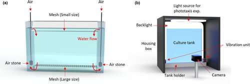
Our platform enables long-term culture and monitoring of daphnid behaviors. (a,b) Schematic illustrating the customized culture and imaging setup. (a) An individual tank. Air stones, connected to the air source, are located in two side columns to create an aerobic environment for daphnids and to separate neonates from mothers via two different sized meshes. (b) Schematic of imaging setup. A tank is set into the imaging setup and recorded via a frontal camera in a computer-controlled environment. An even backlight illumination is used constantly. A housing ceiling light is used for the stimulated phototaxis. The scaffolding ensures an invariant tank placement. Four vibrational motors on both sides of the scaffolding are used for the delivery of vibrational stimulus. Credit: DOI: 10.1111/acel.13571
You recently published a paper in Aging Cell on your Daphnia system. What did you show in this study?
This paper is establishing the baseline for Daphnia as a new model organism for studying aging. We describe the system in detail, including how we set up the tank, fed the animals, removed new offspring, and set the light cycles and temperature. These appear to be boring details, but the whole point is getting the boring details right. We are developing a set of routines that are needed to raise Daphnia in a standardized way that is also scalable.
We also demonstrated the potential of our system for studying aging. When you turn on a light, Daphnia has a reflex to move towards it, and when you turn off the light, they hide. It's a very clear response. In our paper, we show that old Daphnia ignore the light and middle-aged Daphnia have a mixed response: some react, some don't, and some react slowly. This is a nice behavioral assay where our one-minute videos can capture how the reaction to light diminishes with age.
We decided to test metformin, a common diabetes drug that, in the literature, has been shown to extend the life span of worms and flies. We got a beautiful negative result—we conclusively showed that metformin does not affect the life span of Daphnia. We did not necessarily prove or disprove the other papers, but we demonstrated that our system can be used to test a drug of interest and get solid, statistically significant results from a large sample. We also made Daphnia "drunk" by adding ethanol to the tank. Adding 3 percent ethanol had a strong effect on Daphnia behavior, providing proof of concept that our platform can detect the effects of various drugs and interventions.
Your Daphnia system is part of what you call 'radically open, Wikipedia-style science.' What is this concept?
Many people are passionate about aging research. Students ask for projects, volunteers from all over the world write to ask what they can do. There is this huge enthusiasm. These people aren't necessarily experts on aging, but they are hackers, data analysis experts, zoologists, engineers, computer scientists, or even biologists from other disciplines. In building this Daphnia system, I realized that we are very nicely and crisply formulating some bite-sized problems of what has to be done that others can then address. With all of these people willing to help, there is an opportunity to crowdsource some of the science. I began to think about how can we use crowdsourcing to make science more efficient.
Our Daphnia platform is standardized, and consists of a tank that is easy to assemble and animals that are easy to maintain. It is great for educational purposes because people can play and observe. Anyone can do experiments in their lab, no matter where they are in the world. You could do experiments in your basement, test interventions, and immediately upload your one-minute video recordings to a server. With our system, many people can do science, and put the measurements online as soon as they are collected. Some of the measurements will be garbage, but that's how Wikipedia works—it is organized in a way where it's self-correcting. People are going to do crazy stuff with the Daphnia system, but if three teams in three different places repeat the same experiment, it will self-correct.
The idea is that people can assemble the Daphnia system and use it in their own experiments, and they can also improve the system by designing better tanks or developing better machine learning tools to analyze the videos. My job is to develop a cheap, scalable, and reproducible system for Daphnia, and I hope that the system will eventually take off and have a life of its own.
What do you hope to accomplish with your system?
We hope to both screen new drugs and verify drugs reported from research in other organisms. We do not think that any of these drugs are going to substantially extend the life or health of Daphnia. We expect some of them will extend life or health just a little bit. That's why we need thousands of animals and dozens of tanks per experiment: we need a big sample size to reliably find these small differences. Once we find drugs that reliably statistically significantly extend health span a little bit, that gives us the opportunity to ask what are those drugs, what are the targets of those drugs. Then we will know how we should further focus our search.
There are many examples of how research done in mice, or even monkeys, does not translate into humans. On the other hand, there is amazing conservation of processes across species. Metabolism is very conserved—how organisms get energy is largely universal. Many multicellular organisms carry not just similar genes, but similar cell types. We've already found that Daphnia reacts to some drugs developed for humans. If I put a drop of caffeine into the tank, the heart of Daphnia reacts like crazy. Daphnia responds to cardiac drugs, muscle relaxants, and anesthetics. That gives us not just gene or cell type homology, but pharmacological homology. Additionally, the species seem to age in a statistically similar way: after a certain age, the probability of dying for humans doubles every eight years, and in Daphnia it doubles roughly every eight days. Of course, humans are not Daphnia, and there are many things that are not going to translate, but this gives us hope that the processes governing aging in both species are similar.
More information: Yongmin Cho et al, Intelligent high‐throughput intervention testing platform in Daphnia, Aging Cell (2022). DOI: 10.1111/acel.13571


 protein analyses in chronic kidney disease (CKD) and calcific aortic valve disease (CAVD) patients. (A) Spearman correlation between plasma OMD levels and aortic valve calcification (in Agatston scoring units) in CKD patients (n = 65). (B) OMD protein measurements in plasma from CKD patients stratified in groups according to the medial calcification grade/score (CS) of epigastric arteries from these patients (ranging from 0 to 3, where 0 signifies no arterial calcification, 1 and 2 refer to moderate calcification and 3 refers to extensive arterial calcification). Number of patients per group: n = 25 for CS = 0, n = 25 for CS = 1, n = 24 for CS = 2, n = 24 for CS = 3. One-way ANOVA multiple comparison test; data presented as mean with SD. (C) Representative histological images of epigastric arteries from CKD patients with the four different grades of calcification, immunostained for OMD (red signal) and α-SMA (green). Arrows point to OMD positive signal in the tissues. (D) Representative images from consecutive human aortic valve leaflet slides stained with Alizarin red and von Kossa to visualize calcification, or immunostained for α-SMA, OMD and RUNX2. Scale bar as indicated in all images. Insets show corresponding isotype negative control. Differences between groups were considered significant at p-values < .05 (*p < y.05). Credit: DOI: 10.1002/ctm2.682")